Dear visitor,

You may already have seen images of diatoms with their diverse forms and fine structures. Perhaps you also had the opportunity to observe live diatoms under the microscope and noticed that some are capable of moving smoothly or jerky. They often change the direction of movement. The mechanism of movement is not visible.

One of my favourite pastimes is observing diatoms with the focus on species that are motile. I am fascinated by the reaction of diatoms to environmental conditions and external stimuli, such as obstacles or light. In cultures of colonial diatoms, I try to understand the link between motility and the formation of colonies.

I would like to mention that I (Thomas Harbich, see contact) studied physics and spent my working life with typical activities of an engineer in the field of telecommunications and the automotive area.  Since a few years I am retired.

The purpose of this homepage is to encourage you to make your own observations on diatoms. If you like to microscope and follow this suggestion, you will hopefully find useful information on these pages.

You will get information about the following aspects:

    • Cultivation of diatoms
    • Observation and creation of video recordings of the movement of diatoms in different environments
    • Analysis of the resulting images and videos
    • Hypotheses and conclusions from the observations

As there is a comprehensive literature and many web pages to the biology of the diatoms, I restrict myself to some remarks, links and literature references.

From time to time the site will be supplemented by observations and hypotheses. If you are interested in the subject, please feel invited to visit this website now and then. I am grateful for content discussion and suggestions for improvement.

Please note that the content is protected by copyright. If you quote something from this page, please refer to my name and add a link to this page. Please contact me if you want to make use of pictures or videos. I do not pursue any commercial interests.

The site contains many short videos. To view them, a browser is required, which masters HTML5. If a video on a browser is not running correctly, it is recommended to try another browser.

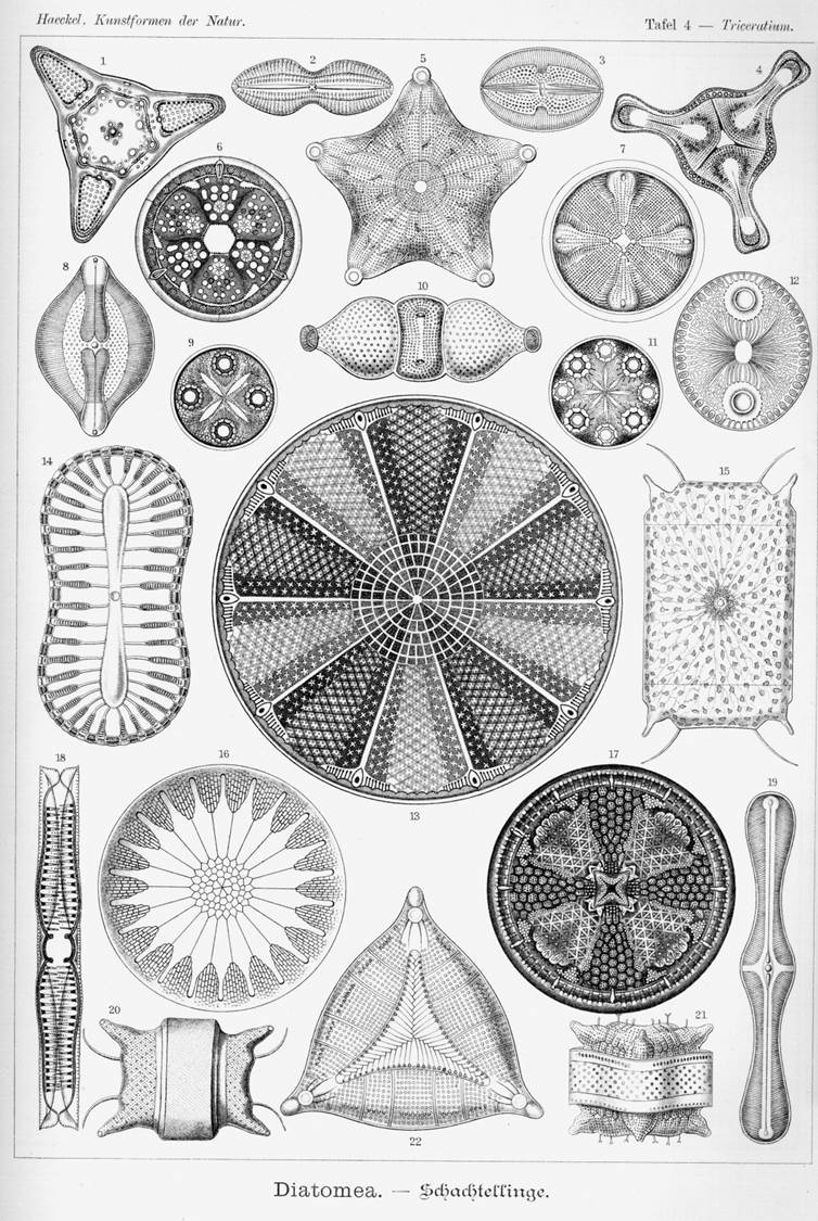
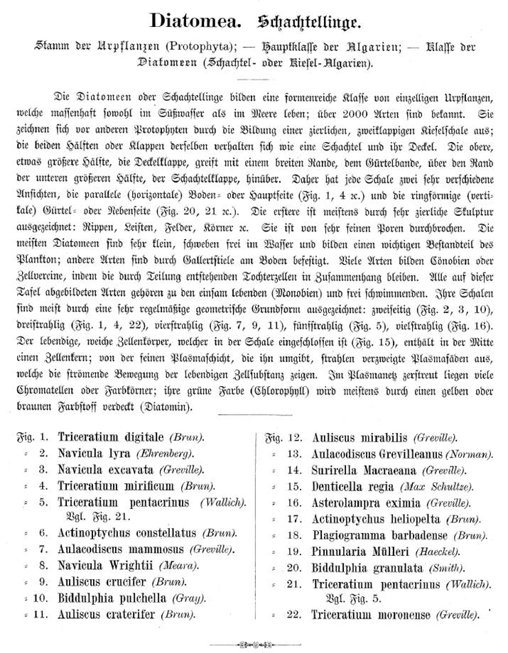
As an introduction to the topic, I show a picture from the book "Kunstformen der Natur" by Ernst Heinrich Philipp August Haeckel (16.02.1834 to 09.08.1919):

Text zur Diatomeenbildseite von Ernst Heinrich Philipp August Haeckel

several diatoms

What are Diatoms?

Let me begin with a few statements about diatoms. In biological terms they form a class of single celled algae. It is characteristic of them that they live in a transparent house made of hydrated silica (SiO₂ + n H₂O), which is coated with an organic material. This exoskeleton (frustule) consists of two halves, the epitheca and the hypotheca. The structure is similar to a petri dish, with the epitheca overlapping the hypotheca. Each theca comprises of a more or less arched valve and the cingulum, a number of associated siliceous bands, the so-called girdle bands (see following picture).

Structure of the diatom
According to their shape, diatoms are divided into centric diatoms that are radially symmetric and pennate diatoms that are bilaterally symmetric. Among the pennate diatoms there are many species which are able to glide over a substratum (see next page).

The size of diatoms ranges typically from a few microns up to about 2 millimetres. However in most cases diatoms are microscopic and require at least a light microscope to observe.

Diatoms are widespread and can be found in almost all fresh and saline waters like brooks, rivers, lakes and sea. Even moist soil serves as a possible habitat. Some diatoms are floating, others live at the bottom of a body of water.

Diatoms show an immense variety of shapes and structures. Their casing exhibit pores allowing them to exchange nutrient and waste. The valves of some diatoms have a slit, the so called raphe allowing them to move over surfaces of grains of sand, stones or the surface of aquatic plants.

Like all plants diatoms use photosynthesis to gain solar energy. Apart from chlorophyll a and c, fucoxanthin serves as photosynthetic pigment which gives diatoms a golden-brown colour.

There is a huge taxonomic diversity with hundreds of diatom genera. One finds different estimates on the number of recent species. It may be 100,000 or even more.

Diatoms reproduce by asexual (vegetative) and sexual reproduction. When a cell divides (mitotic division), a smaller valve is re-formed, so that after the division one has one cell of the same size and a smaller one:

mitosis


The following picture illustrates the vegetative reproduction over 5 generations:
asexual reproduction

Statistically, therefore, the size decreases (MacDonald-Pfitzer rule). When the smallest diatom reaches a minimal size, sexual reproduction is required to gain a cell of maximal size. In rare cases, there is also a vegetative cell enlargement.

The sexual reproduction of pennate diatoms has a great variety of variants. Diatoms are diploid, so they have a double chromosome set. By reduction division (meiosis) one or two haploid gametes are formed in each diatom, that is to say, germ cells with a simple chromosome set. In pennate diatoms isogamy prevails, in which the gametes are of the same size and are not flagellated. By fusion of the gametes a zygote or two zygotes with a double chromosome set are produced. Finally, each zygote grows to the auxospore and forms a new vegetative cell (initial cell) with two valves, which has the maximal size.

It is also possible that two gametes of the same gametangium will fuse into a zygote, from which the auxospore and the initial cell are formed. This self-fertilization is called automixis.

Furthermore, it was observed that an auxospore can mature without reduction division (asexual reproduction). In addition conjugation was discovered in pennate diatoms.

For details on the steps, see Round et. al. (2007). Terminology and pictures can be found in Irena Kaczmarska et. Al (2013).


F. E. Round; R. M. Crawford; D. G. Mann (2007), Diatoms: Biology and Morphology of the Genera, Cambridge University Press; 1 edition (2007)

Irena Kaczmarska , Aloisie Poulíčková , Shinya Sato , Mark B. Edlund , Masahiko Idei , Tsuyoshi Watanabe & David G. Mann (2013): Proposals for a terminology for diatom sexual reproduction, auxospores and resting stages, Diatom Research, DOI:10.1080/0269249X.2013.791344

Links

Useful links to English sites:Link icon

Wikipedia, Diatom https://en.wikipedia.org/wiki/Diatom
International Society for Diatom Research: http://www.isdr.org/
University College London, Micropalaeontology Unit, Diatoms: http://www.ucl.ac.uk/GeolSci/micropal/diatom.html
The Phycology Section: Ecology and Taxonomy of Freshwater Algae, particularly Diatoms: http://diatom.ansp.org/

 
Databases, Diatom Identification:

Diatoms of the United States: https://westerndiatoms.colorado.edu/
Algae World: diatoms: http://rbg-web2.rbge.org.uk/algae/diatoms.htm
Introduction to Diatom Identification: http://rbg-web2.rbge.org.uk/ADIAC/intro/intro.htm


Useful links to German sites:

Wikipedia, Kieselalgen: https://de.wikipedia.org/wiki/Kieselalgen
Diatomeen – Kurzeinführung: http://www.diatomeen.de/
Diatomeen-Homepage von Dr. phil. nat. E. Alles: http://www.kieselalgen.com/JSIndex.html


Books

The following compilation is a tip to the very extensive literature.
Further references to publications are given in the appropriate place.


Books in English:

The Diatoms: Biology and Morphology of the Genera
F. E. Round; R. M. Crawford; D. G. Mann
Cambridge University Press; 1 edition (2007)

The Biology of Diatoms (Botanical Monographs)
Dietrich Werner (Editor)
University of California Press, Berkeley, California (1977)

Identification of Freshwater Diatoms from Live Material
E.J. Cox
Springer (1996)

Algal Culturing Techniques
Robert A. Andersen (Editor)
Academic Press (2005)


Books in German:

Diatomeen im Süßwasser-Benthos von Mitteleuropa : Bestimmungsflora Kieselalgen für die ökologische Praxis;
über 700 der häufigsten Arten und ihre Ökologie
Horst Lange-Bertalot (Editor)
Koeltz Scientific Books

Kieselalgen in Binnengewässern
Lothar Kalbe
VerlagsKG Wolf (2005)

Kieselalgen : Biologie, Baupläne d. Zellwand, Untersuchungsmethoden
Kurt Krammer
Kosmos Verlags-GmbH (1990)

Algenreinkulturen, ihre Herstellung und Erhaltung
E. G. Pringsheim
Jena Fischer (1954)


Articles relating to the homepage:

Harbich, T. (2021), On the Size Sequence of Diatoms in Clonal Chains. In Diatom Morphogenesis (Diatoms: Biology diatom morphogenesisand Applications) Vadim V. Annenkov (Editor), Richard Gordon (Editor), Joseph Seckbach (Editor), Wiley-Scrivener; First published: 29 October 2021, https://doi.org/10.1002/9781119488170.ch3


Harbich, T. (2021) Some Observations of Movements of Pennate Diatoms in Cultures and Their Possible Interpretation. In Diatom Gliding Motility (Diatoms: Biology and Applications) S.A. Cohn (Editor), K.M. Manoylov (Editor) and R. Gordon (Editor), Wiley-Scrivener, Beverly, MA, USA; First published: 20 July 2021, https://doi.org/10.1002/9781119526483.ch1

The sections are:

  • Kinematics and Analysis of Trajectories in Pennate Diatoms with Almost Straight Raphe along the Apical Axis
  • Curvature of the Trajectory at the Reversal Points
  • Movement of Diatoms in and on Biofilms
  • Movement on the Water Surface
  • Formation of flat Colonies in Cymbella lanceolata


Please also have a look at these publications:
diatom gliding motility

Alicea, B., Gordon, R., Harbich, T., Singh, U., Singh, A., & Varma, V. (2021) Towards a Digital Diatom: image processing and deep learning analysis of Bacillaria paradoxa dynamic morphology. In Diatom Gliding Motility (Diatoms: Biology and Applications) S.A. Cohn (Editor), K.M. Manoylov (Editor) and R. Gordon (Editor), Wiley-Scrivener, Beverly, MA, USA; First published: 20 July 2021, https://doi.org/10.1002/9781119526483.ch10

Preprint: https://www.biorxiv.org/content/10.1101/2019.12.21.885897v2


Gebeshuber, I. C., Zischka, F., Kratochvil, H., Noll, A., Gordon, R., & Harbich, T. (2021), (2021) Diatom Triboacoustics. In Diatom Gliding Motility (Diatoms: Biology and Applications) S.A. Cohn (Editor), K.M. Manoylov (Editor) and R. Gordon (Editor), Wiley-Scrivener, Beverly, MA, USA; First published: 20 July 2021, https://doi.org/10.1002/9781119526483.ch11


 Cymbella spec. in dark field (40x time lapse) Visualization of the motion of the diatoms from the video by calculating the maximum over all frames (click to enlarge)


Movement

A number of benthic species have the ability to move. Nearly all of them have a raphe. On smooth ground they glide in straight or curved paths, the shape of the paths depends on the curvature of the raphe. They also show complex movements such as sudden reversing, turning around the apical axis, erecting, horizontal rotation about one point of the cell, pirouettes in the erected state, etc. Overall, the movements seem to be randomly and it is not necessarily clear what benefits are generated.

Possible benefits are in particular:

  • Optimization of light conditions, because many motile species show positive or negative phototaxis. A photophobic response can also be observed in which diatoms react strongly on local changes in the light intensity with reversal of the direction of motion.
  • Periodic vertical migration of diatoms inhabiting sand deposits in particular in intertidal zones. These sediments can be disturbed by tides and currents (see review article Harper (1977))
  • Search for places with better nutrient concentration or other favorable chemical environmental conditions (chemotaxis). In the publication of Karen Grace V. Bondoc et al (2016) it is shown that Seminavis robusta moves towards a silicate source.
  • Colonization of new habitats
  • Search and approach to a partner for sexual reproduction. The structure and function of sexual pheromones has been elucidated for Seminavis robusta (Frenkel, Johannes. PhD Thesis (2014) and Bondoc et al. (2016))
  • Leaving the copulation envelope in certain species (see post about sexual reproduction)

An approach to the target of the movement achieves a diatom by varying the movement activity, in particular by controlling the duration of the movement in one direction.

At this point I would like to make a comment from my point of view. It is common to all mentioned advantages of the movement that they are due to a change of location. When you look at the movement of some species, you can doubt whether this can always be the motivation. Cymatopleura elliptica usually rotates slowly around a vertical axis, interacts uncontrolled with the environment and comes hardly from the spot (see video). In many cases, the benefit of mobility is only temporary, as in sexual reproduction. Nevertheless, the majority of the diatoms in a culture is independent of their size always in motion. In certain cases, the benefits could also have a physiological background such as the regulation of energy balance.

Since the discovery of the movement of diatoms, one struggles for an understanding of the mechanism of motion. As early as 1838, Ehrenberg described a snail-like foot for creeping (Die Infusionsthierchen als vollkommene 0rganismen. 1838 p. 175), which was not proved to be correct. Up to this day, there are numerous theories; none of them is definitively confirmed. They range from a movement due to capillary effect to a cytoplasm flow and to the recoil principle. Today a secretion of mucilage through the raphe and a movement of the mucilage along the raphe are usually used as an explanation, whereby muscle proteins serve as a drive. The prevailing hypothesis is given by Edgar, L.A. & Pickett-Heaps J.D. (1984). A compact representation was created by Menzel, D. and O. Vugrek (1997) and is reproduced below with the original illustration and translated text:


actin myosin mechanism

Schematic representation of the sliding motion in patented diatoms.
(a) Bottom of a migrating cell. A mucilage trail is formed from the posterior raphe.
(b) cross-section along the line in (a). The silicified cell wall consists of two parts (blue-gray), which, like the two halves of a petri dish, are on top of each other. The chloroplasts are shown in green; the nucleus in the center of the cell is omitted for the sake of clarity. Underneath the raphe is a pair of actin filaments (blue), which are used as pathways for the transport of mucilage vesicles (pink). The vesicles fuse at the ends of the raphes with the plasma membrane. Each vesicle unloads a mucilage trail (red) to the outer side of the membrane where it swells and is pressed through the gap of the raphe. One end of the thread adheres to the substrate, the other remains bound to the cell membrane, and it is assumed that this end is connected with a motor molecule (dark violet), which transports the thread along the actin bundles. Upper and lower raphe produce mucus filaments at the same time.
(c) detail of b.
Menzel, D. and O. Vugrek, Muskelproteine in Pflanzenzellen. Biologie in unserer Zeit, 1997. 27(3): p. 195-203. Copyright Wiley-VCH Verlag GmbH & Co. KGaA. Reproduced with permission.


Two known problems of this theory are mentioned here:

  • The movement of myosin on actin filaments always takes place in one direction. The movement of the diatoms, however, changes the direction of motion, with no difference in speed. Diatoms have one or two raphe systems on one valve. Each raphe system allows movements in both directions. In some species one can even observe that particles are transported in the opposite direction along the same raphe and collide (Nultsch, W. (1962)).
  • The raphe of many motile diatoms is not simply an open cleft. Some move in spite of an almost closed tongue in groove slit. Other mobile species do this by means of a canal raphe, which is located on the valves and is connected to the interior of the cell only by a series of pores.

At least amendments are necessary to explain the observations by the described actin-myosin transport.

J. Wang, S. Cao, C. Du and D. Chen have investigated the movement at Navicula sp. and propose an updated model. In this model the diatoms are moved by pseudopods protruding out of the valves. One can not necessarily assume that this model can be applied to other genera as well.

On this homepage, observations will be added to the already existing countless ones on the movement behaviour of diatoms. According to my means, these are always observations with the light microscope and one can ask what is still to be observed at all after 200 years of light microscopic examination. In fact, much is being reproduced. Other observations perhaps encourage the reader to his own observations. In several cases I could not check how far these are described in the literature. I would be grateful for hints. Such observations cannot yield a new explanation of the mechanism of motion but may allow a critical look at the described model conception.


Edgar, L.A. & Pickett-Heaps J.D. (1984), Diatom locomotion., Progress in Phycological Research Vol. 3: 47-88

Frenkel, Johannes. PhD Thesis (2014). Struktur und Funktion von Sexualpheromonen der Diatomee Seminavis robusta. Friedrich-Schiller-Universität Jena, Biologisch-Pharmazeutische Fakultät

Harper, M.A. (1977). Movements. In: The Biology of Diatoms, (D. Werner, ed). 224-249, Blackwell, Oxford

Karen Grace V. Bondoc, Jan Heuschele, Jeroen Gillard, Wim Vyverman & Georg Pohnert. Selective silicate-directed motility in diatoms. Nature Communications http://dx.doi.org/10.1038/ncomms10540

Bondoc, Karen Grace & Lembke, Christine & Vyverman, Wim & Pohnert, Georg. (2016). Searching for a Mate: Pheromone-Directed Movement of the Benthic Diatom Seminavis robusta. Microbial Ecology. 72. 10.1007/s00248-016-0796-7.


See also:

Jeroen Gillard, Johannes Frenkel, Valerie Devos, Koen Sabbe, Carsten Paul, Martin Rempt, Dirk Inzé, Georg Pohnert, Marnik Vuylsteke, Wim Vyverman: Metabolomik unterstützt die Strukturaufklärung eines Sexual­phe­romons von Kieselalgen., Angewandte Chemie, DOI: 10.1002/ange.201208175

Nultsch, W. (1962) Über das Bewegungsverhalten der Diatomeen., Planta 58: 22.

Wang, J., Cao, S., Du, C. & Chen, D. Underwater locomotion strategy by a benthic penate diatom Navicula sp. Protoplasm 250, 1203–1212 (2013).


Colonial Cymbella cistula attaced to the substrate by mucilage stalks

(30x time lapse)

 Craticula cuspidata in darkfield and brightfield (20x time-lapse)


Purpose of cultivation

Why is it useful to cultivate diatoms that you want to observe? From our point of view, the advantages are as follows:

  • Compared to an observation on a fresh sample, the genus and possibly the species can be identified with greater certainty and assigned to the observation.
  • The identification can take place independent of the observation.
  • The use of just one species is prerequisite for the reproducibility of experiments.
  • A large number of diatoms of a certain species is available. This allows investigations in which a statistical statement is the goal, such as in the case of population dynamics.
  • Other organisms and impurities do not interfere with the observations (see videos below).

Thus, the conditions under which one observes become well controllable. The development of a population can be investigated as a function of external parameters such as temperature, light conditions or composition of the culture medium.

Moreover, the culture vessel (e.g. petri dish) itself can be used for observation.

A sample from the river Neckar. Most striking are colonies of the diatom Bacillaria paxillifera (Bacillaria paradoxa). These colonies exhibit a unique movement based on the motility of the diatoms relative to the neighbouring diatoms. The observation is particularly affected by detritus. (4x time lapse). View into a culture that was created with Bacillaria paxillifera from the Neckar river. The quality and the possibilities for observation are significantly improved compared to the observation in the sample. ( 4x time lapse).


On the pages on the subject of cultures we briefly describe what Mr. Kurt Schneider and I can convey from our experience. On this occasion, I would like to thank Mr. Oliver Skibbe (http://www.larger-than-life.de/micropage/start.html) for his valuable advice.


Cookies make it easier for me to provide you with my services. With the usage of my services you permit me to use cookies.