
")


Quantitative analysis of directly attached Cymbella colonies
Growth of culture
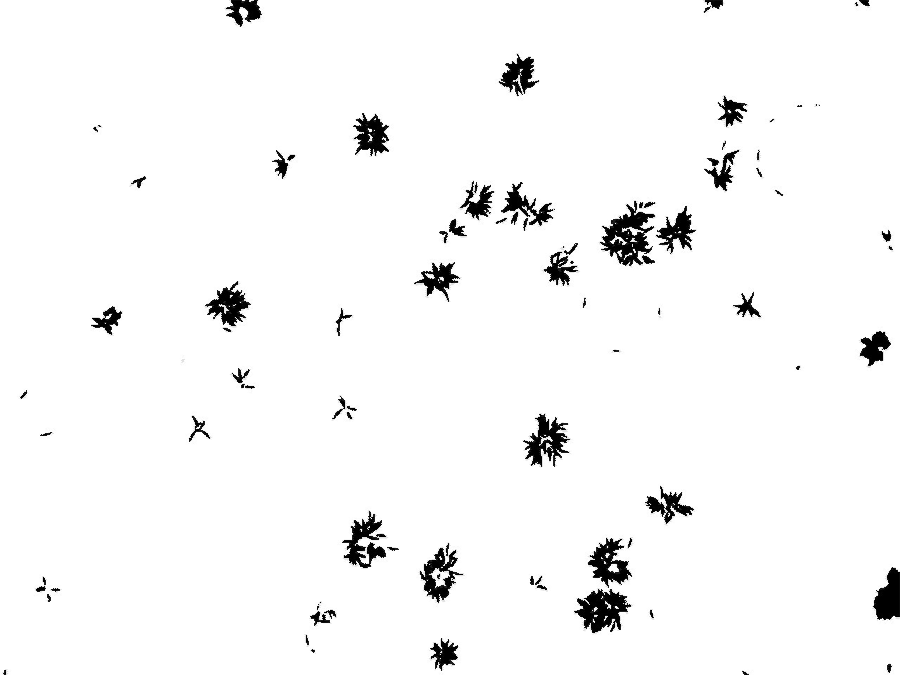
In order to quantify the growth of the culture and colonies, the area of the diatoms in the visible region was determined. At first, the number of images was reduced to 60 images per day. This selection of images is analyzed as a stack of images using ImageJ (Fiji). After conversion to grey tones and setting a threshold value, a binary representation is obtained in which the diatoms appear black on white. The Particle Analysis (‘Analyze Particles’) now calculates the size of the contiguous image parts in number of pixels for each image, which can be output in the form of a csv-table. Therefore, Excel is the appropriate tool for further evaluation and graphical presentation.
At first one would assume that the total area occupied by diatoms increases steadily with the development. The diatoms, which are released from the colonies at brightness, reduce the area of the colonies accordingly. In return, the diatoms between the colonies increase the total area. However, there are various effects that lead to fluctuations which are correlated with the light-dark cycle. Diatoms in colonies can overlap in the image. Furthermore, the area of a diatom in the image depends on its inclination towards the substrate. This inclination varies on average depending on the position in the colony. The number of diatoms in a colony is therefore only roughly proportional to the area taken. Apparently, the surfaces of the individual diatoms located outside the colonies are surprisingly small, as can be seen from the explicit comparison of images in darkness and subsequent brightness. This may be due to errors caused by the low resolution or effects of threshold value calculation. Therefore, the surfaces were divided into colonies and individual diatoms in an image. The classification has been validated by exemplary counting:
- Areas smaller than a lower threshold of a few pixels (about 1 to 4 pixels) are sorted out. They are caused by small particles on the substrate and irregular boundaries
- All objects larger than an upper threshold (in this evaluation 70 pixels) are considered colonies.
- All objects in between are mainly characterized by individual diatoms. It is possible, for example, that two diatoms overlap in the image and, due to their projection, occupy a small area so that they are counted as one cell, although they represent either two diatoms or a very small colony. However, this is rarely the case.
In rough approximation, the number of diatoms in the colonies can be estimated by dividing the total area taken by them by an average area of diatoms in a colony. This conversion factor must be determined appropriately.
The following figure shows the number of individual diatoms (blue) and the number of diatoms in colonies (red) over time. The phases of bright light are marked with a yellow bar.

With the start of the light phase the number of individual diatoms increases rapidly. It falls off again in the course of the light phase. The area of the colonies decreases in mirror image. The conversion factor was selected in such a way that the sum of the two curves shows the smallest possible fluctuations with the light-dark cycle. This is not a precise quantitative count of the number of diatoms in the colonies. The following figure below shows the total number of diatoms resulting from the sum of the two curves discussed. An exponential function (red line) can be easily adjusted:

The corresponding logarithmic representation shows strong fluctuations at the beginning of cultivation, as there are only a few diatoms in the observed range (see note above):

Movement activity
The number of diatoms between the colonies can be determined by classifying the sizes of the components of an image. However, this does not correspond to the number of diatoms that move, because some diatoms remain in place, especially at low brightness. In order to estimate the number of moving diatoms, the particle analysis plugin can be used well again. Diatoms of the genus Cymbella have a characteristic speed in their movement. If one superimposes two images whose recording times differ so much that moving diatoms have covered a distance of at least their own length, then they can be seen twice in this picture.
 For superimposition one uses the calculation of the minimum offered by the Image Calculator. In the next step, the number of individual diatoms in the superposition image and in the single image is determined by classifying the size. The difference between the two numbers results in the number of diatoms shown twice, i.e. the number of moving diatoms. In the image on the left, even 6 binary images were superimposed, each of which was taken at intervals of one minute. The animated picture shows such superimpositions in succession during a light phase. In order to determine the number of moving diatoms, the difference between the number of individual diatoms in the superimposed image and the number of individual diatoms in the non-superimposed image is divided by 5 since the moving diatoms additionally appear 5 times. Overall, a period of 5 minutes is used to evaluate the movement activity, during which the size of the colonies does not change significantly. The following picture shows the movement activity of the diatoms over the last 10 days of the observation time:

Apparently, the movement activity falls off quickly after the light phase has set in. However, this is just one example. Cymbella species can be observed that show high activity throughout the light phase. The already shown video on the activity in light and dark phase demonstrates this well.
It can be assumed that there is a strong dependence on light intensity. It is sufficient to bring the culture under the strong illumination of the inverse microscope to activate the diatoms at any time. As has already been shown, the colonies can be dissolved to a considerable extent at high light intensities.
The described procedure for determining the activity is reasonably easy to carry out, but it is also error-prone. Sometimes diatoms stop or resume their movement during the period of superposition. Diatoms can enter or leave the observed area. The colonies also change their shape to a certain extent through the movement of diatoms. Especially at high densities, images of diatoms may overlap with images of other diatoms that were there at another time. In the present case of small diatoms and a large observed region, the difficulty lies mainly in the quality of the binary images. This is expressed in imprecise classification. Nevertheless, I believe that the procedure is a simple and good approach, especially in comparison with the tedious manual counting process.