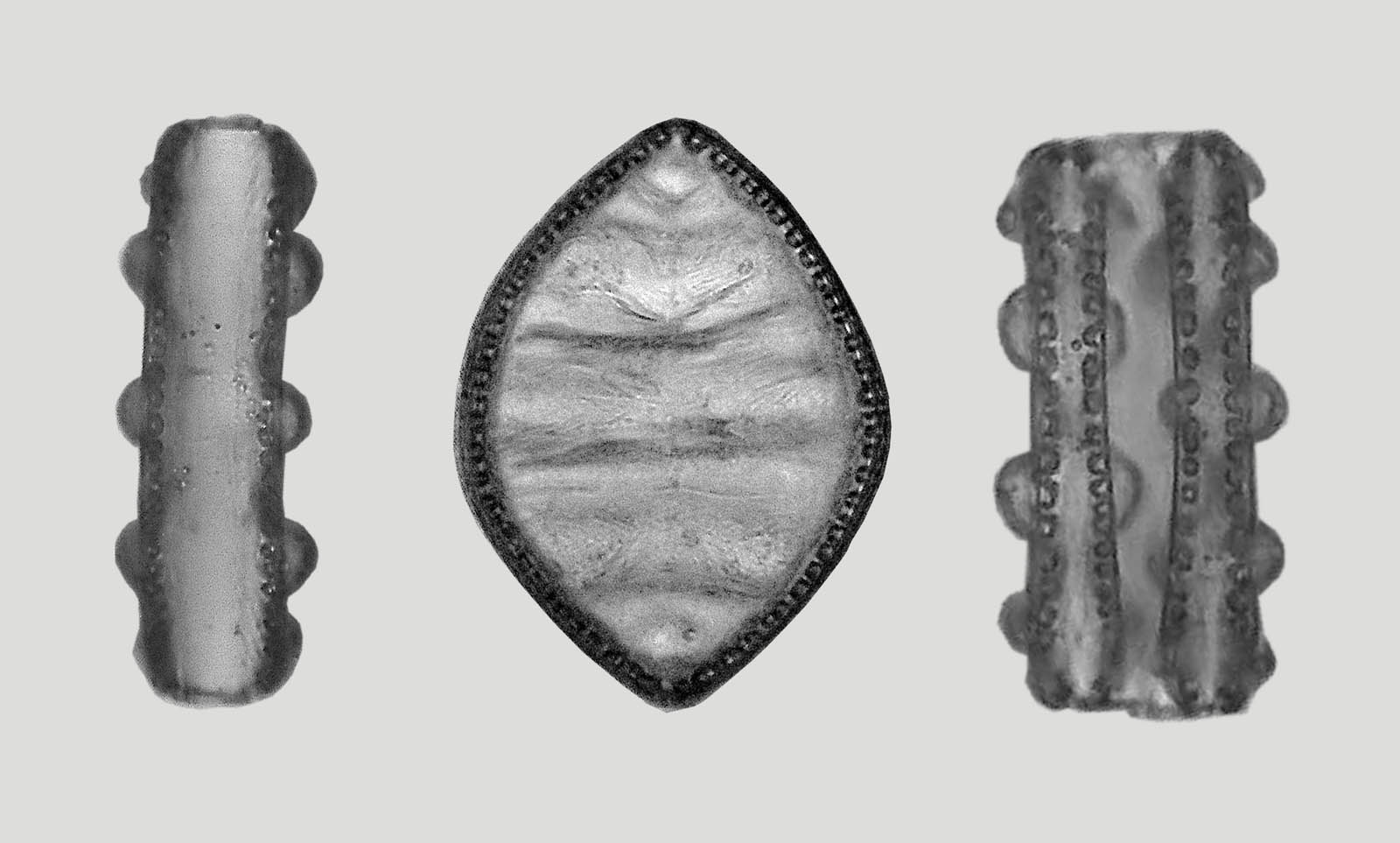
Images of Cymatopleura elliptica after annealing
The species Cymatopleura elliptica shows a very characteristic appearance in valve view and belt view. Below you can see some diatoms of this species in both views. The diatom on the right side of the picture is currently in division. The images were composed of image stacks and placed in front of a homogeneous background.
It is difficult to gain a spatial impression from the projections into a plane. This is easier if the diatoms are located by chance in a position that lies between valve view and belt view. Such a situation is found in this image:
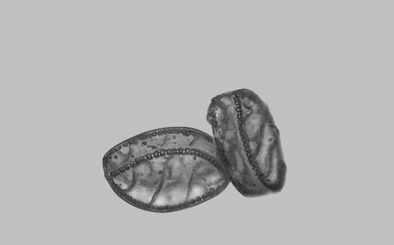
This image was also created using a stack of images, with the focus planes approximately equidistantly spaced. Therefore, a depth map of the captured structures can be calculated. This is accomplished by the software (freeware) "Picolay" from Heribert Cypionka (https://www.picolay.de/). The following animated gif image was created with it, which shows the view of the right and left eye alternately.