Introductory words and pictures of chain-forming diatoms
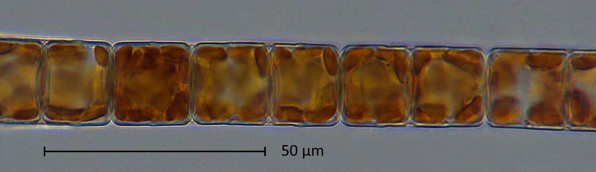
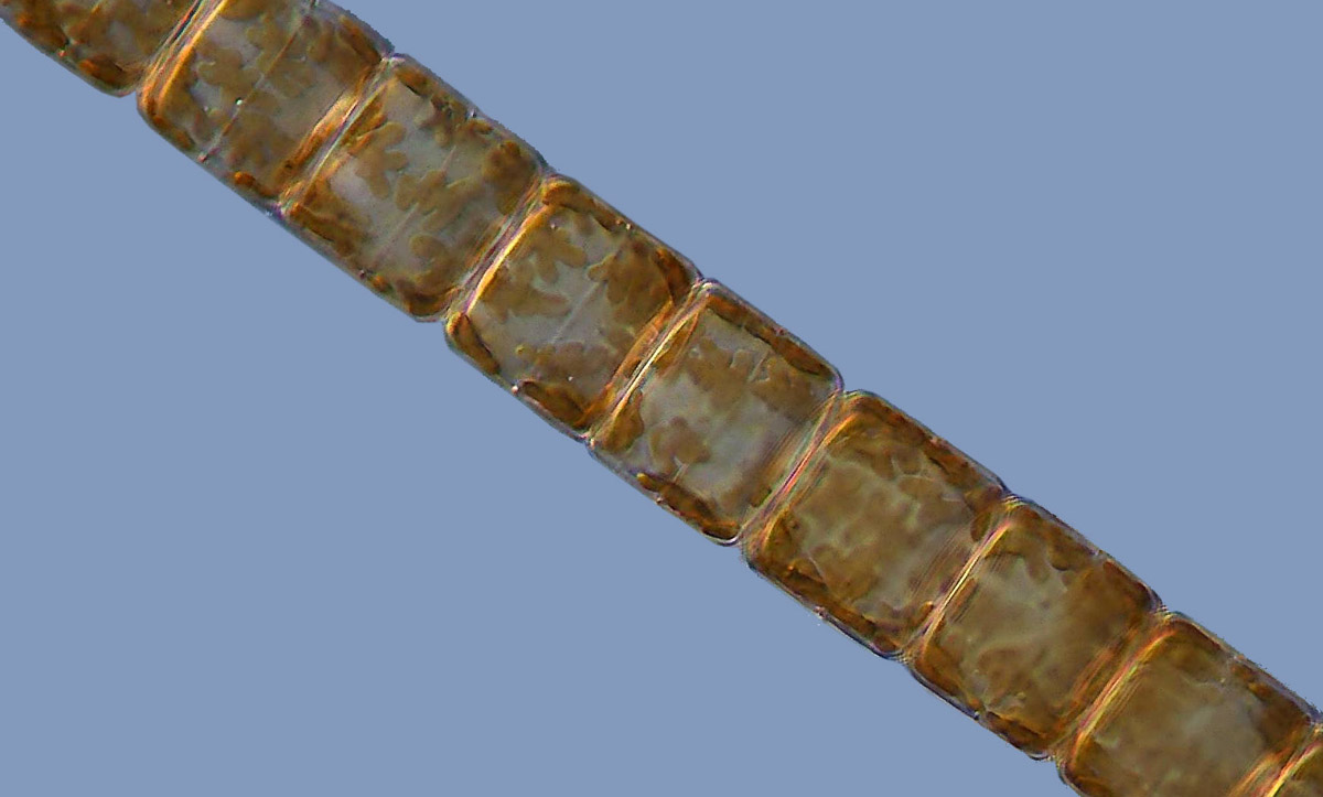
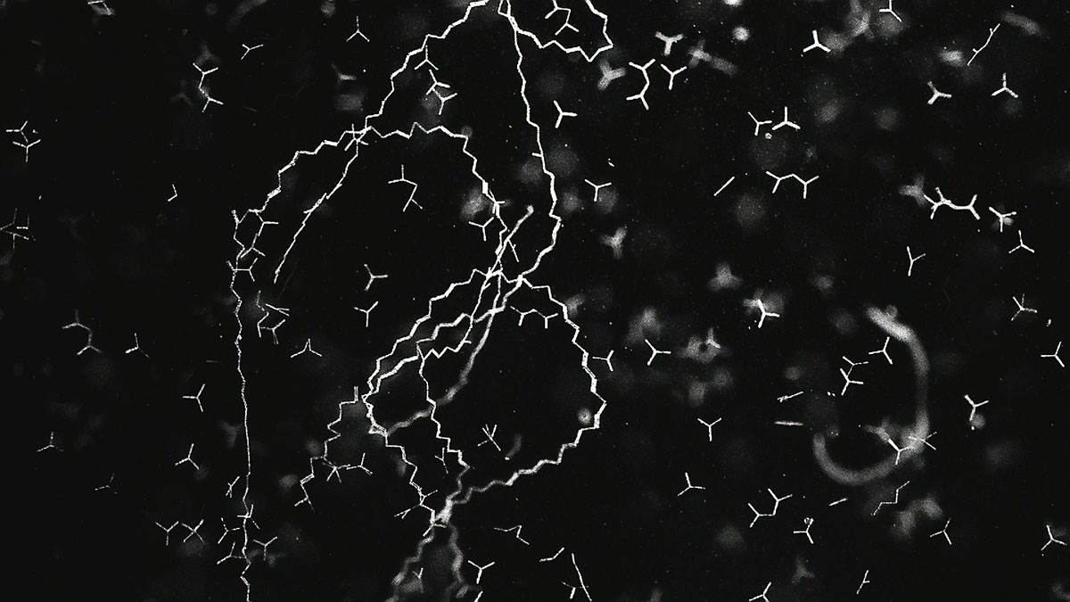
Some diatoms do not separate after an asexual reproduction, but adhere together and form chain-like colonies. These colonies can take the form of filaments, ribbons, stars or fans. The picture above shows a section of a colony of Melosira varians (click to enlarge). In cultures one can often observe such colonies consisting of thousands of connected diatoms.
This page is intended to display images of colony-forming diatoms that have been kept in culture.
One example is a species of the genus Gomphonema. A video recording of this motile diatom can be seen in this video in 8-fold time-lapse. The Diatoms can detach from colonies and form a new colony elsewhere. Especially in newly established batch cultures, one can observe how it moves among the still-young colonies.
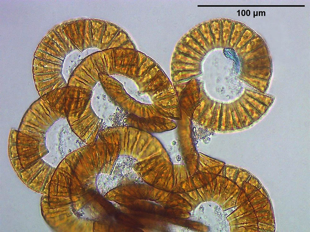
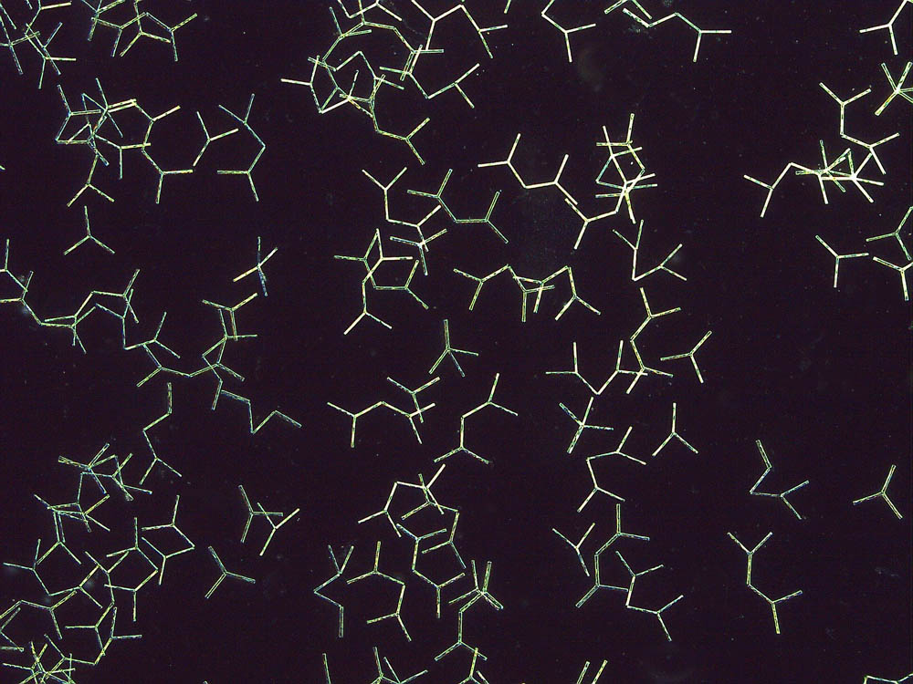
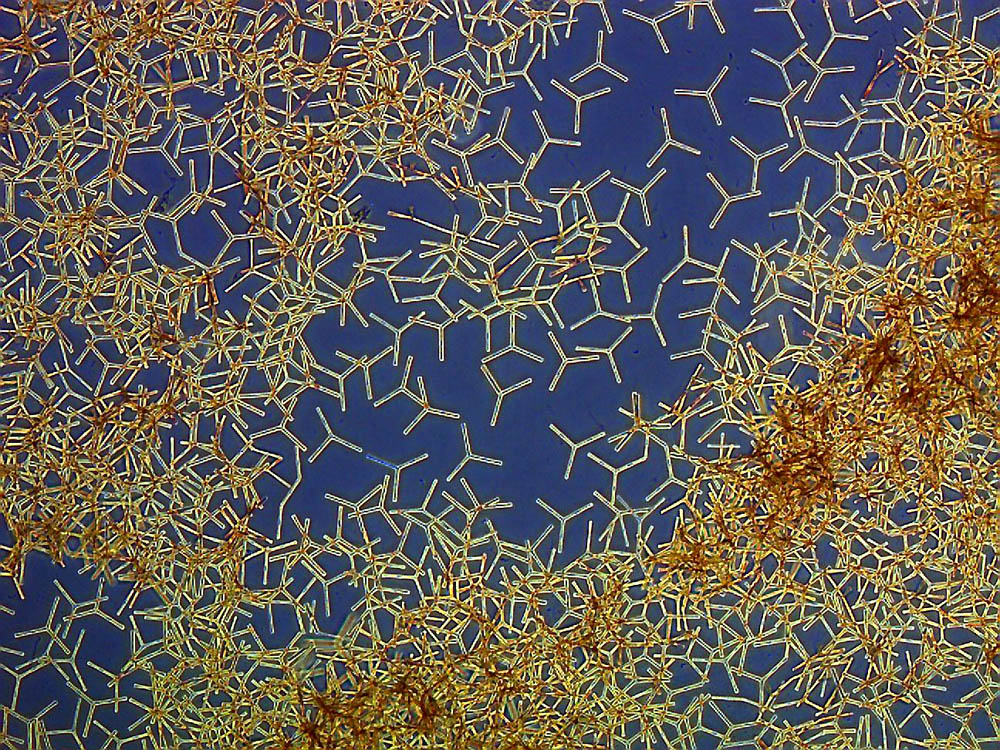
In the subsequent image gallery you can see colonies of Gomphonema sp.forming overlapping fans. Gomphonema sp. is a motile diatom.
Click on the thumbnail to enlarge the image.
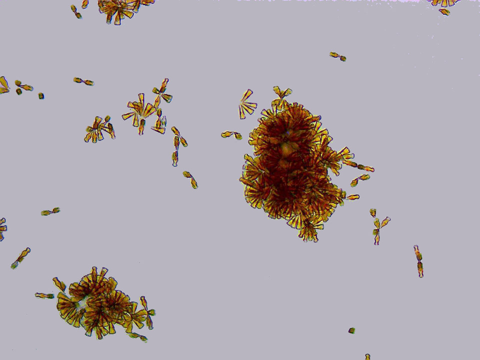
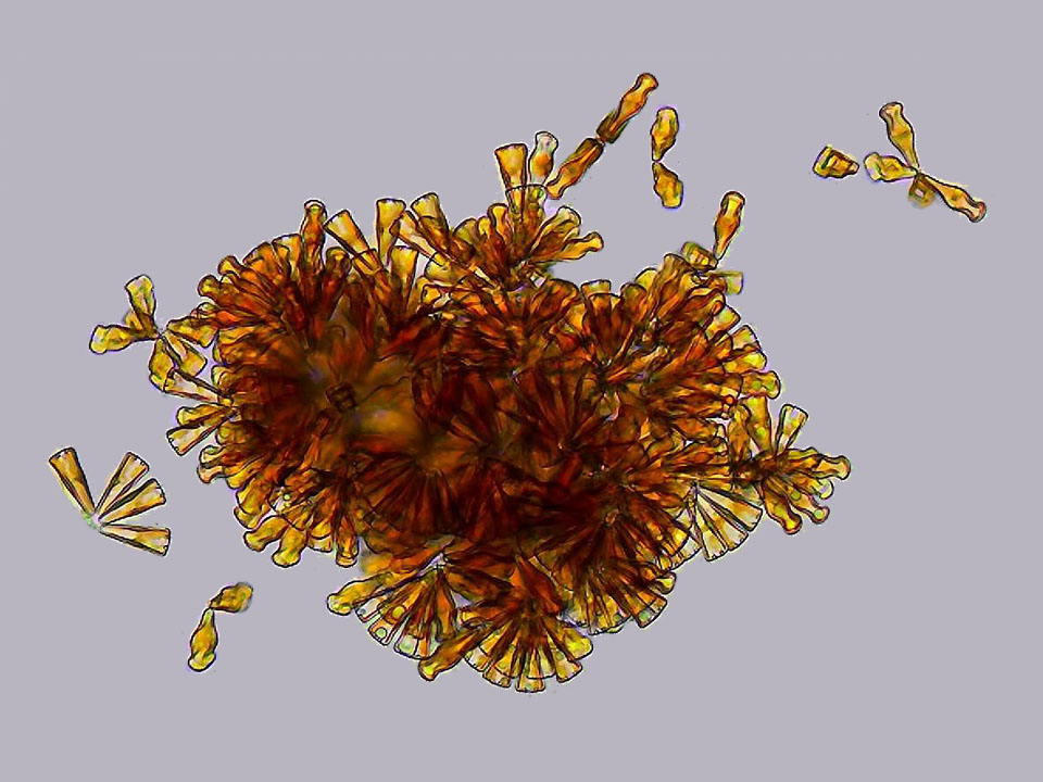
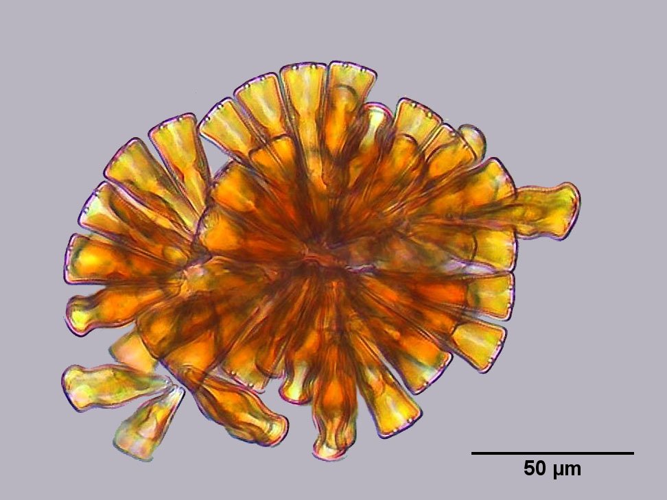
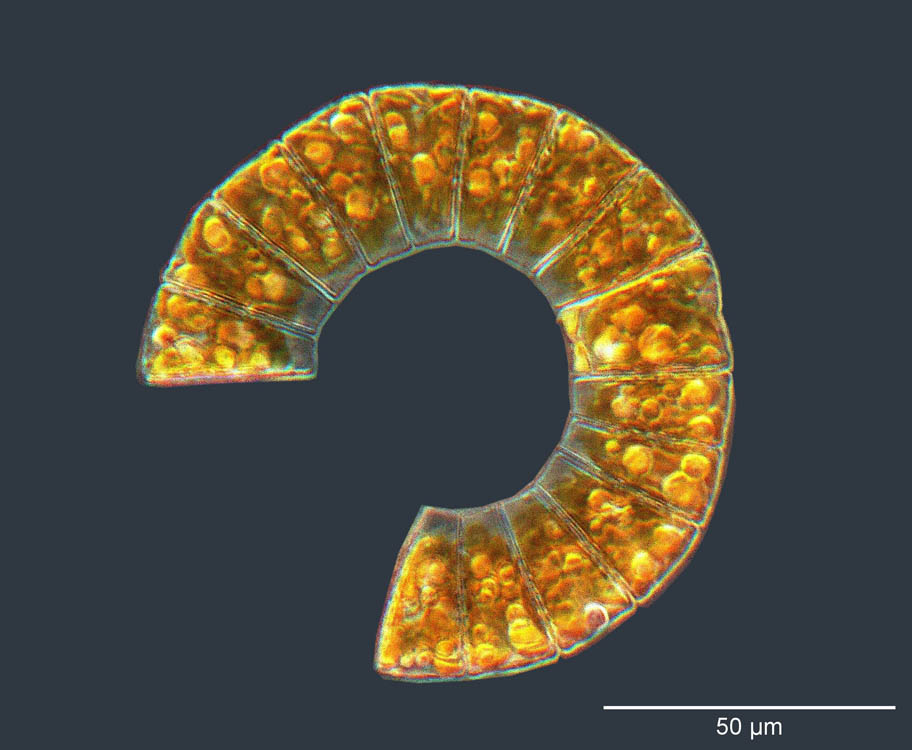
Meridion circulare also forms fan-shaped colonies, which can be seen next.
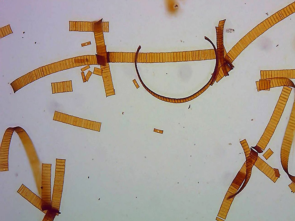
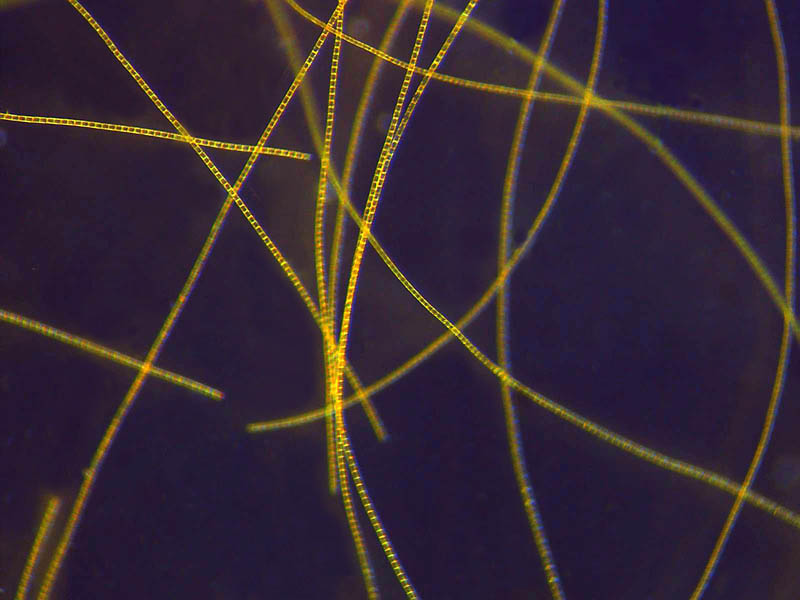
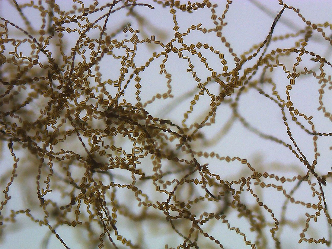
In the following series of images you see an Eunotia sp. culture in which long ribbon-shaped chains are formed. Eunotia is also motile. In another section there is a contribution on its complex movement patterns.
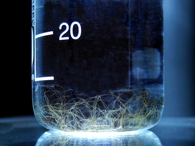
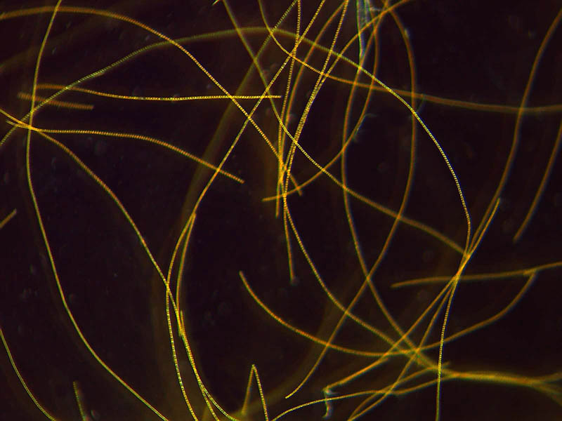
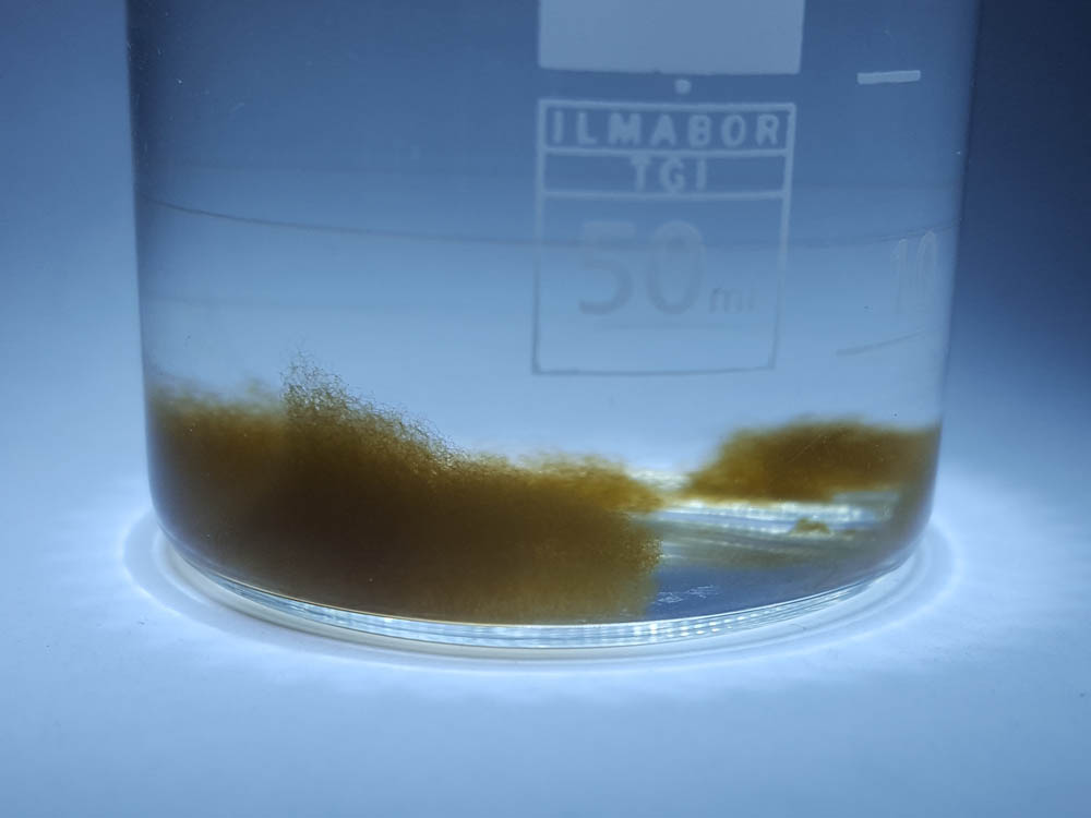
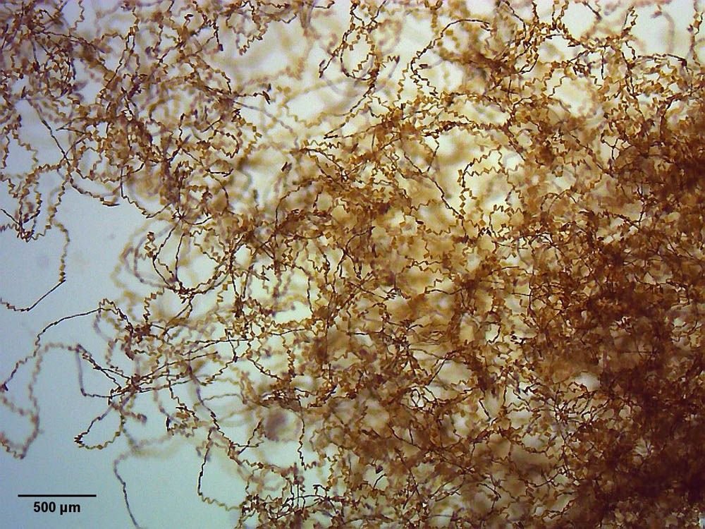
Below you find images of Melosira varians. First you see a small beaker containing nutrient solution, into which a few fragments of a short M. varians colony have been given. Already after a few days in suitable lighting, a fine web of long colonies appeared. This web was taken with the stereo microscope at two magnifications. In order to achieve sufficient contrast, the photos were taken with dark field illumination. The original colony has already been broken into several fragments.

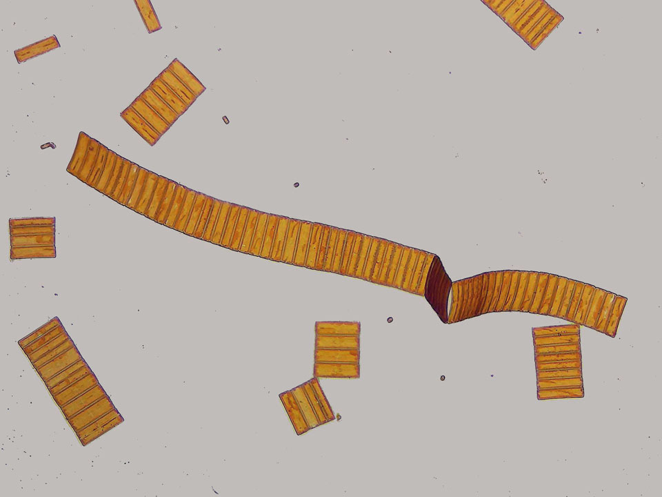
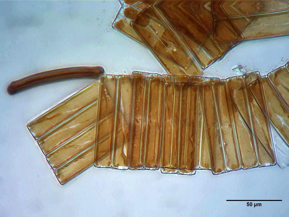
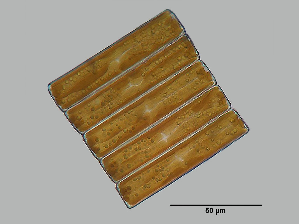
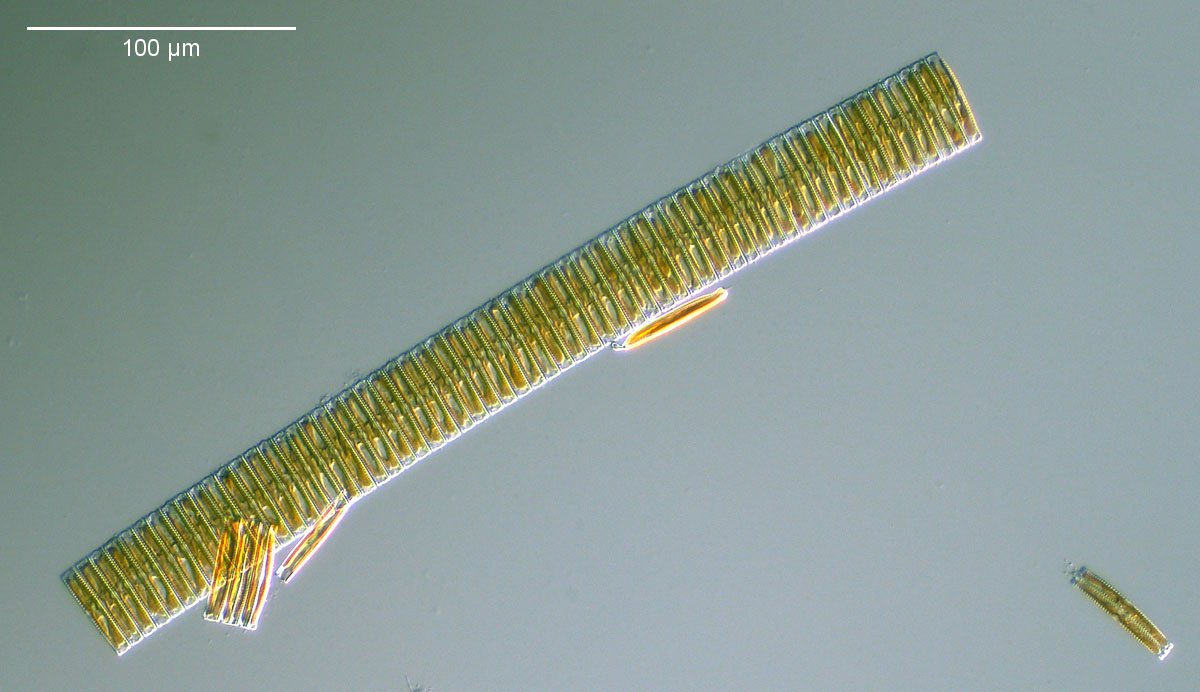
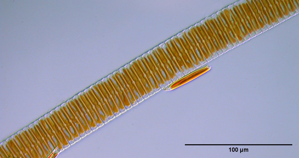
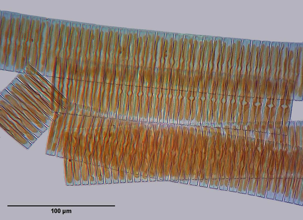
Below are images of a Fragilaria sp. culture.
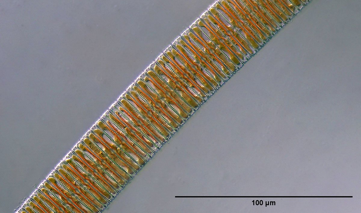
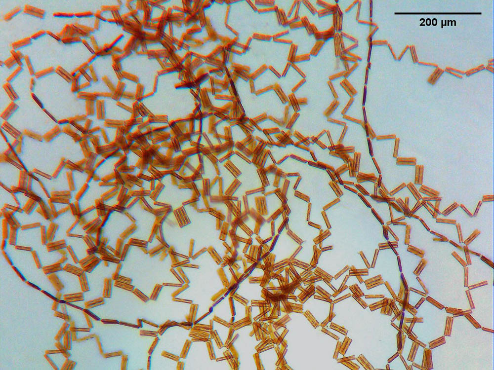
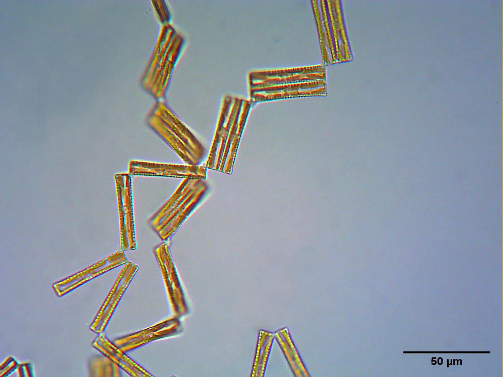
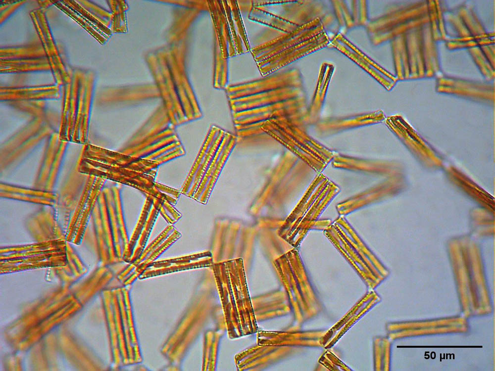
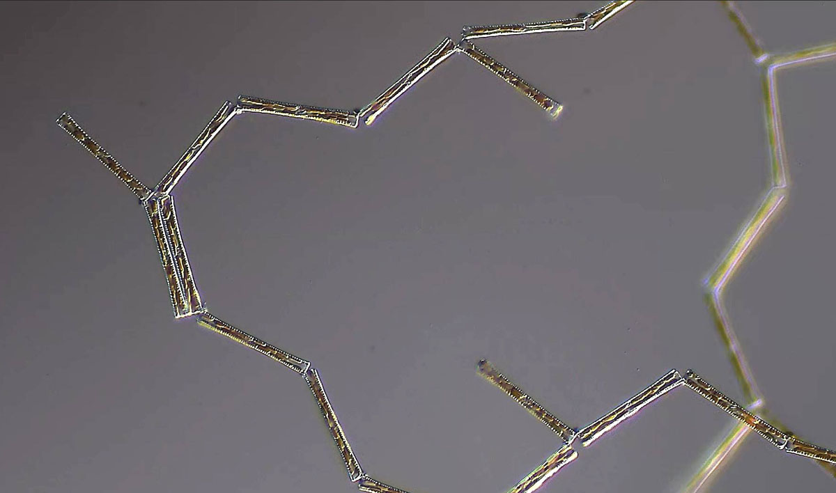
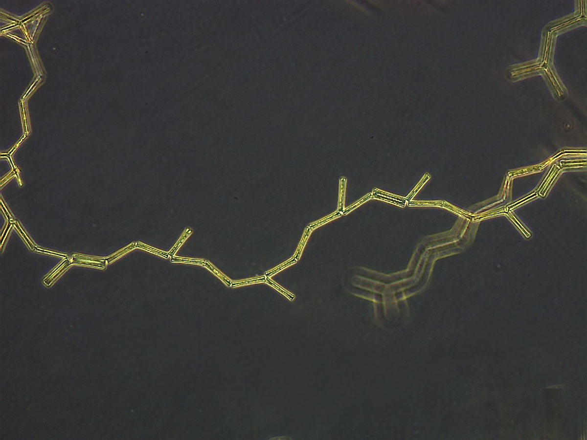
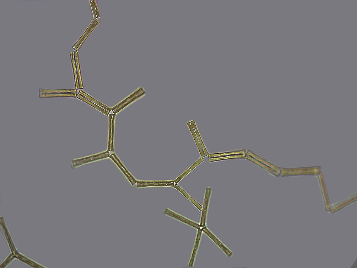
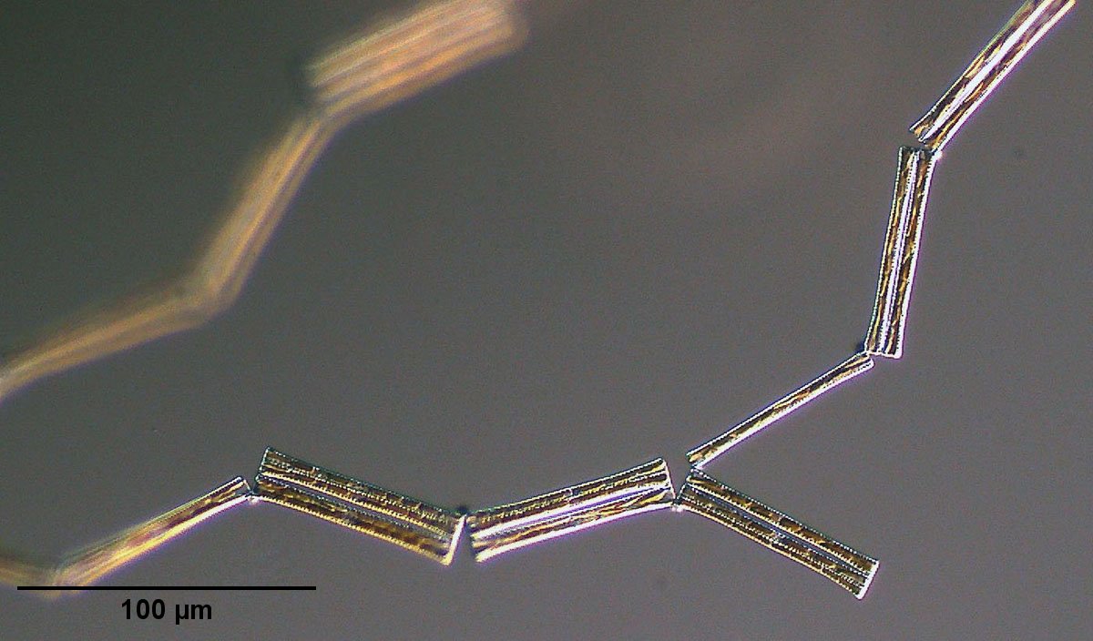
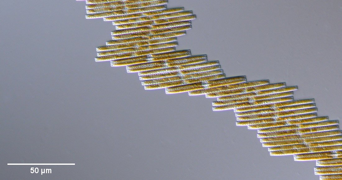
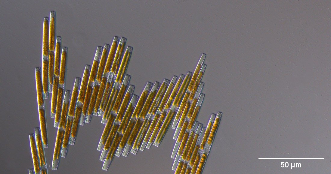
Images of Diatoma vulgaris in culture follow. The diatoms do not lie parallel to each other, because they usually do not separate completely, so that zigzag forms develop. In the DIC images, the ESP pads connecting the diatoms are clearly visible.
An image gallery of a Diatoma culture of unclear species is shown below. This diatom also quickly develops a clump of connected diatoms in a beaker with nutrient solution.
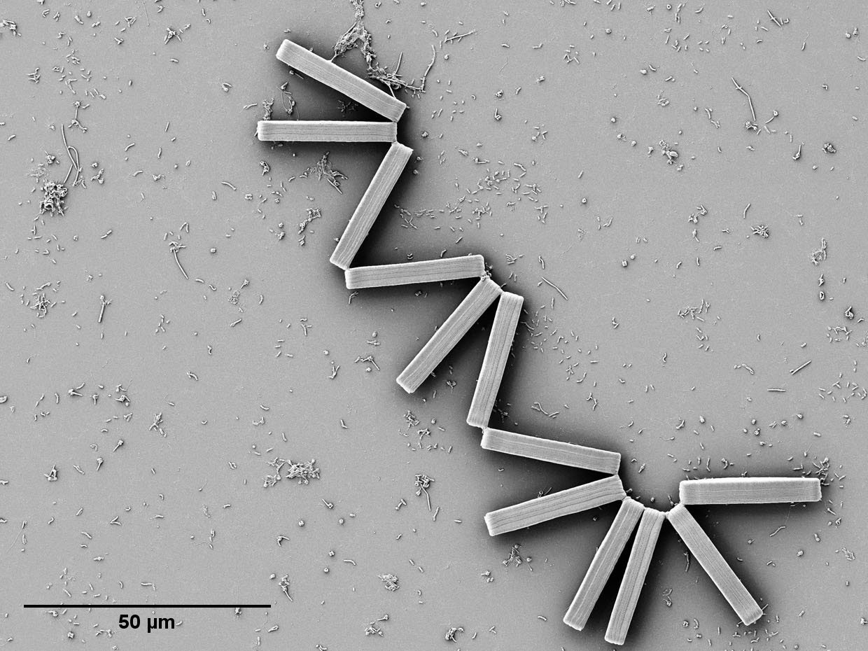
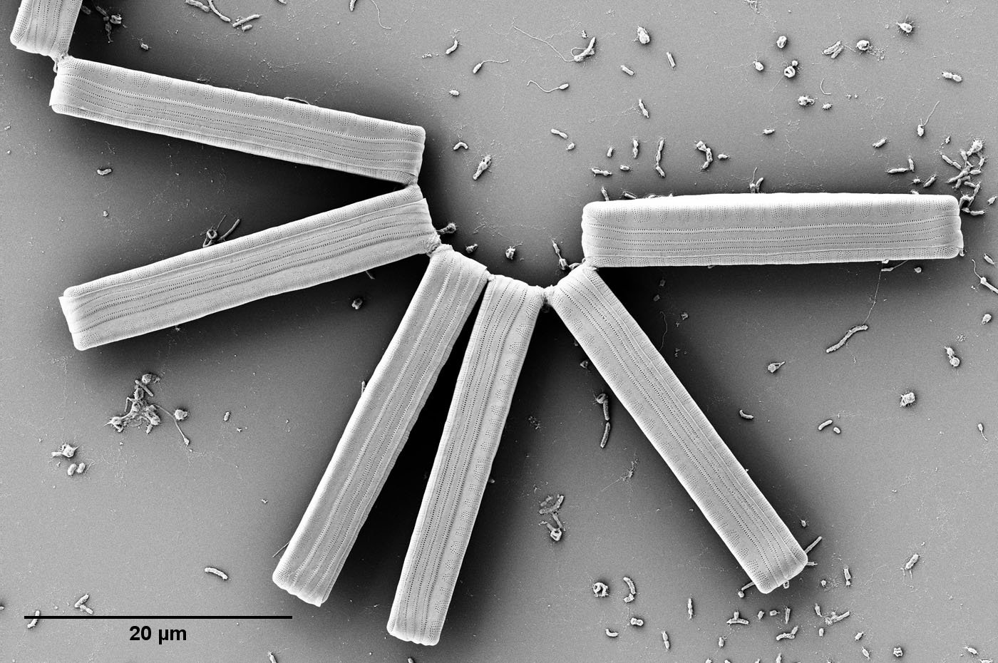
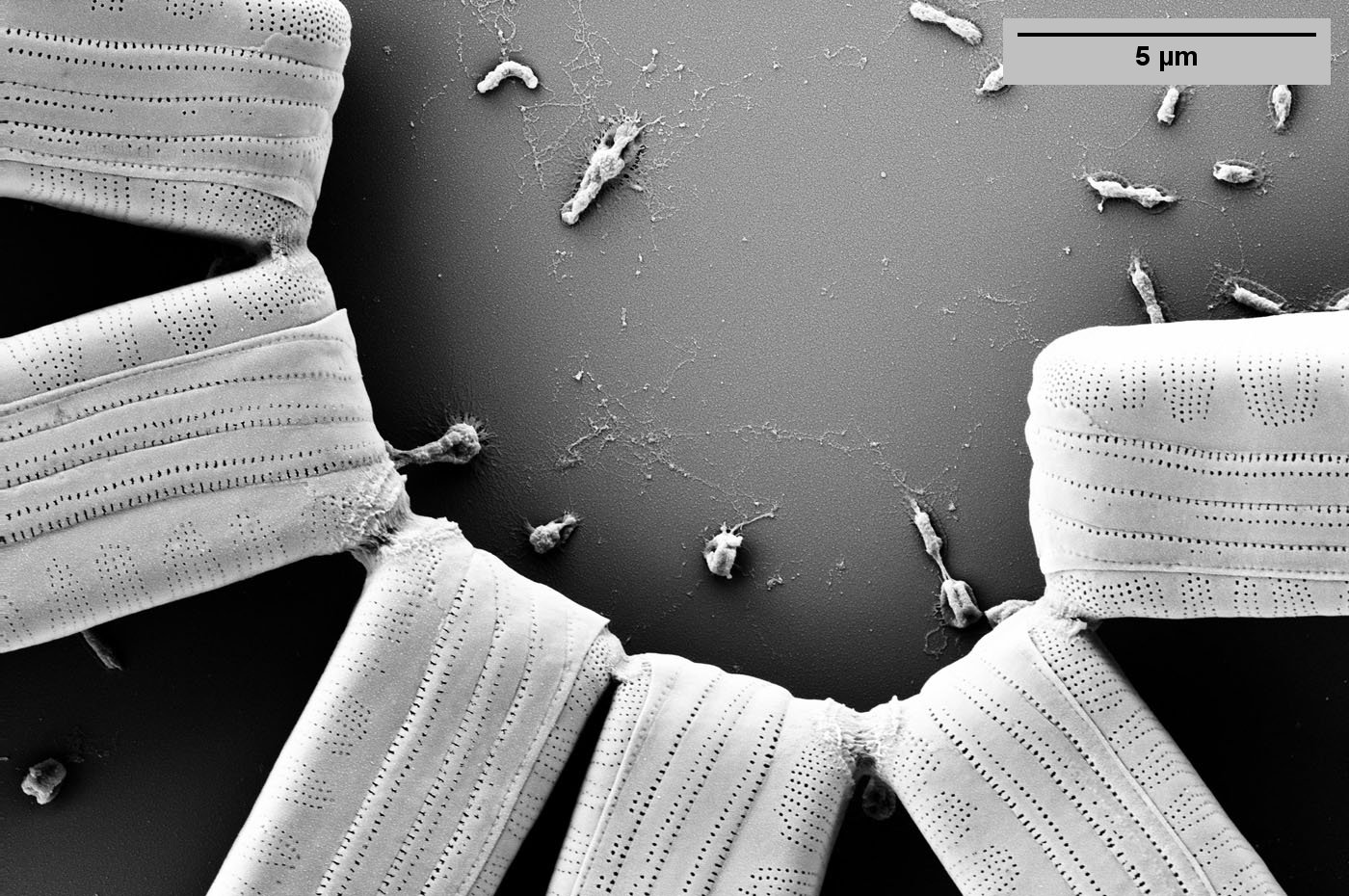
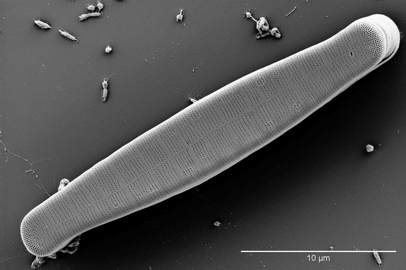
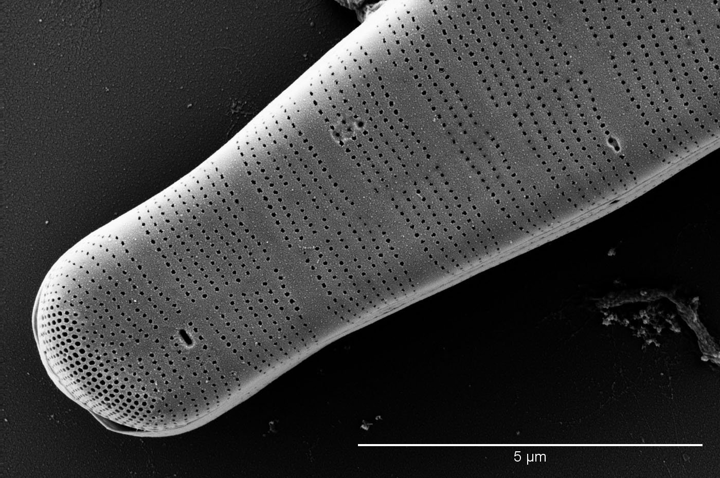
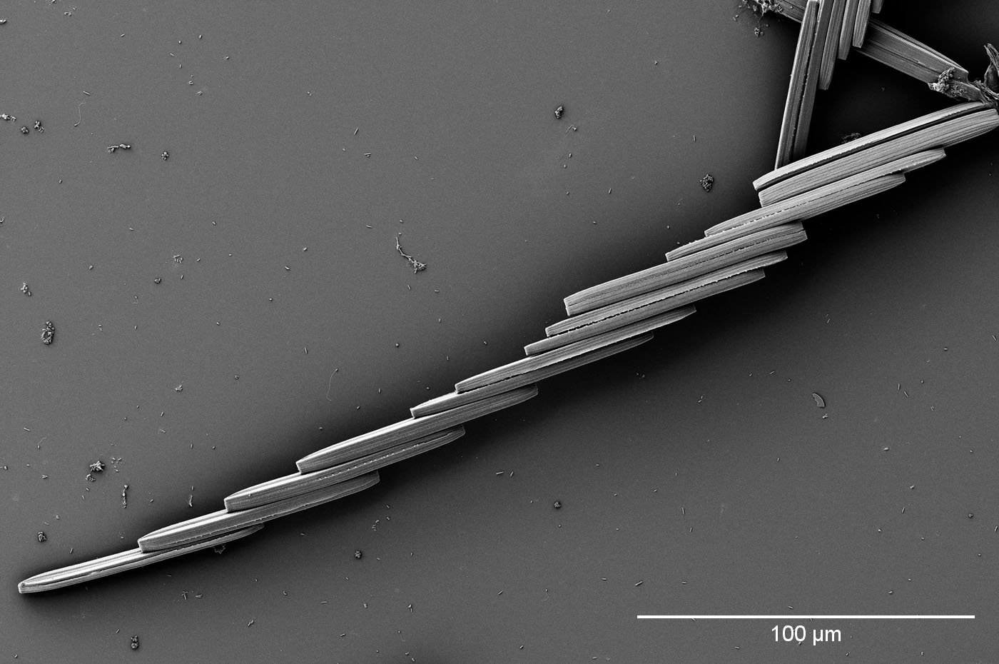
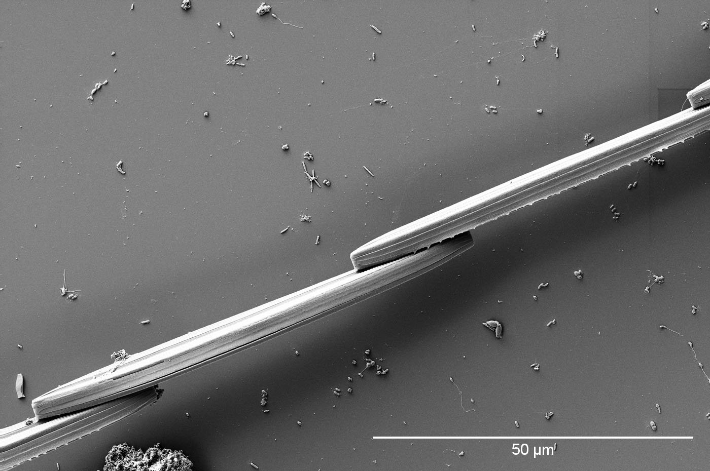
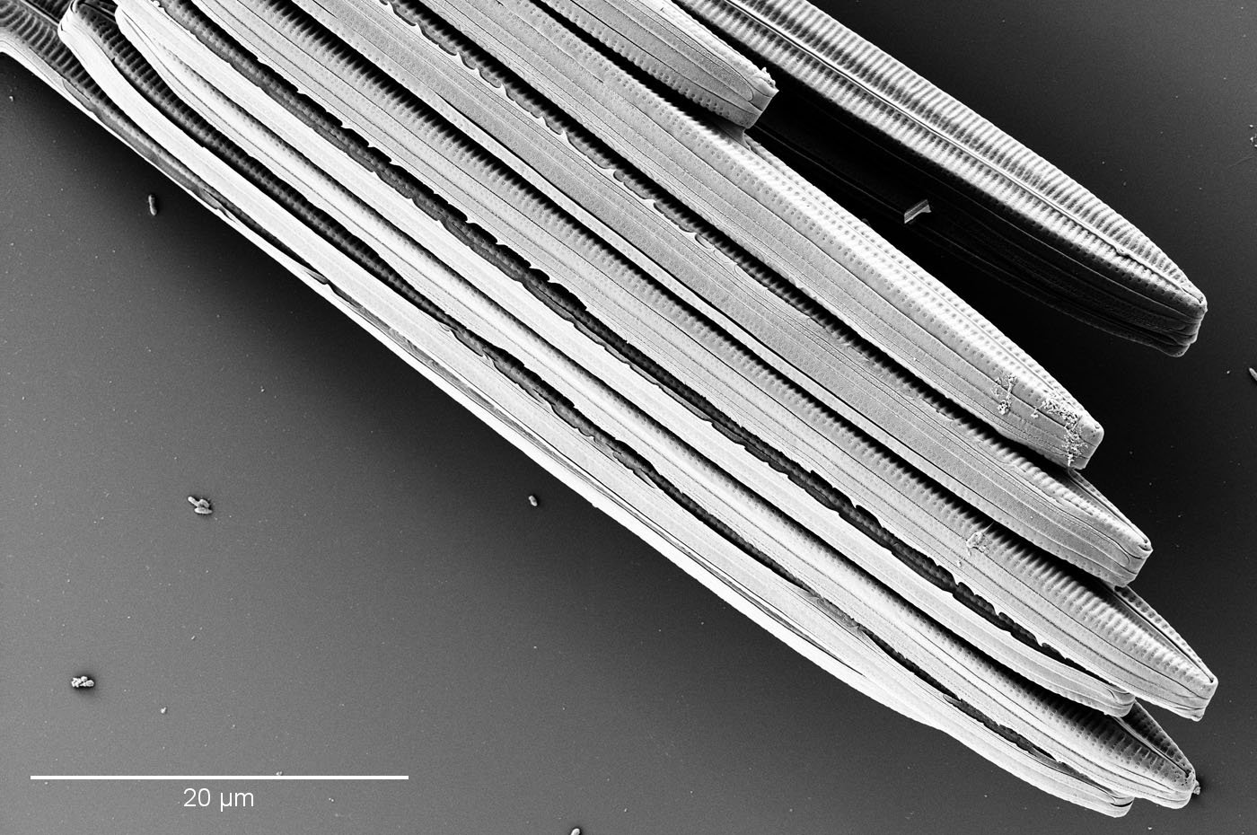
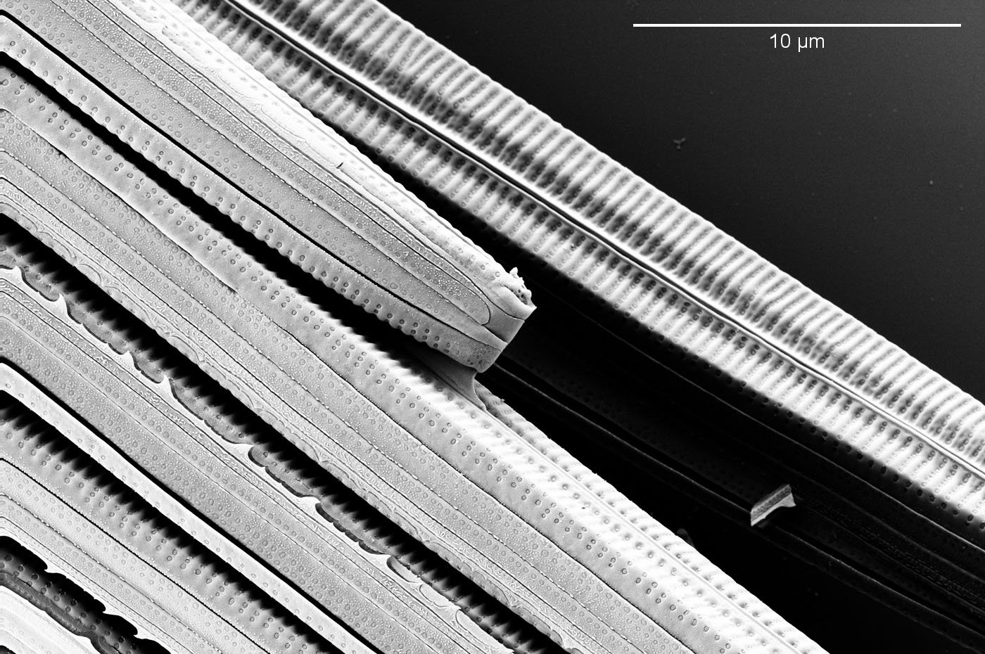
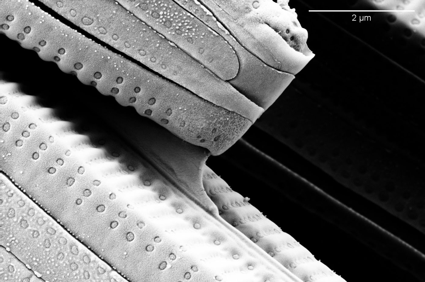
SEM images have been taken for this Diatoma strain. Before inoculating a new culture, small round coverslips were placed into the Petri dish, which were then colonized by the diatoms. The diatoms on the cover glass were fixed with glutaraldehyde, rinsed with distilled water and dehydrated in baths with increasing isopropanol concentration. The SEM images were kindly produced by Dr. Wilfried Nisch, NMI Reutlingen, https://www.nmi.de/en/. On the images (picture gallery below right) you can see well the extracellular polymeric substances (EPS), which connect the diatoms to a chain near the apices.
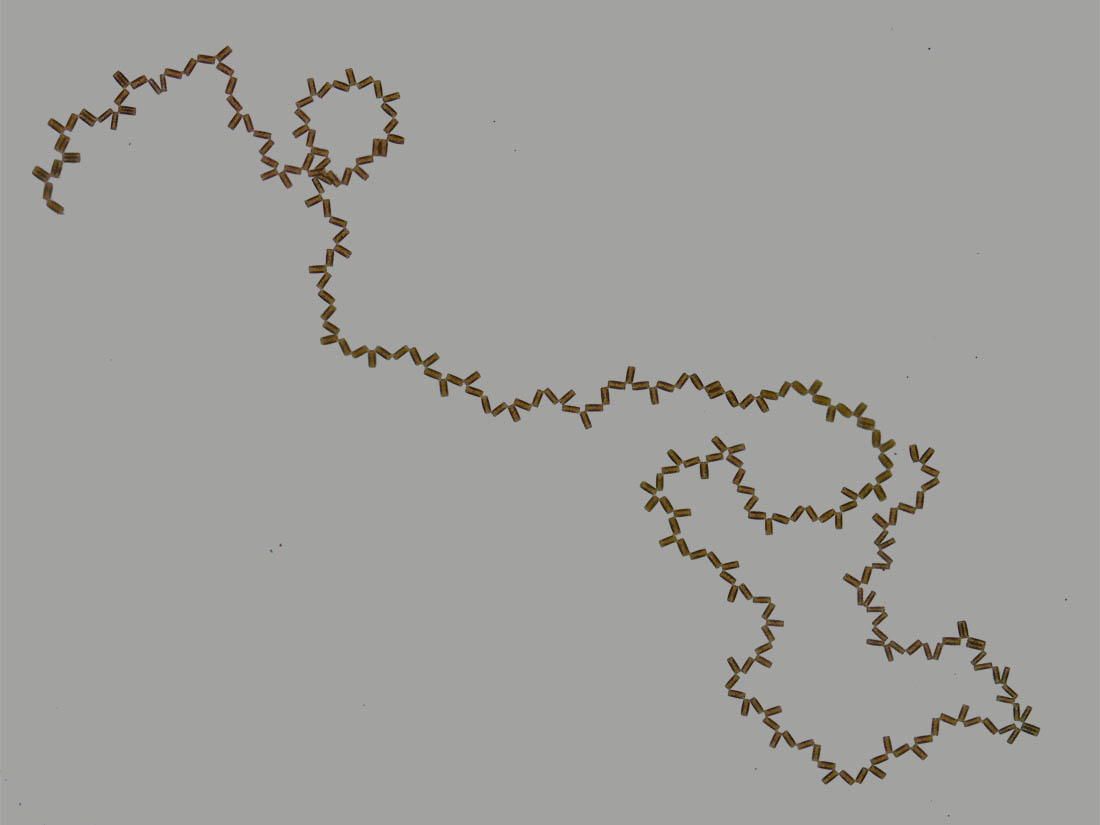
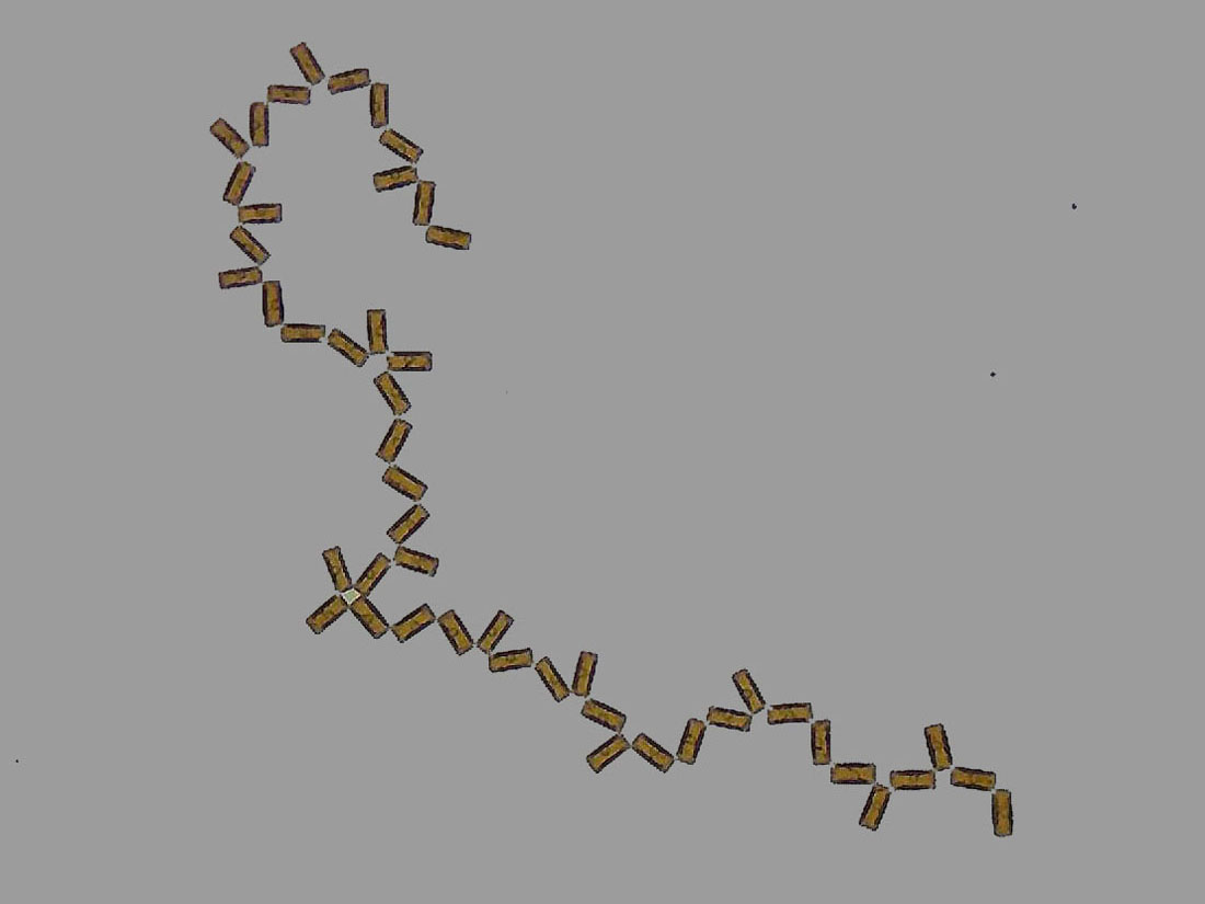
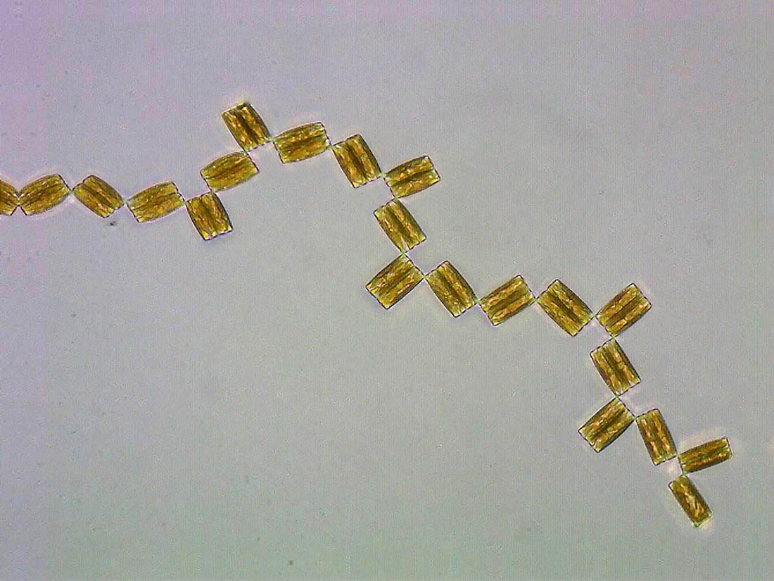
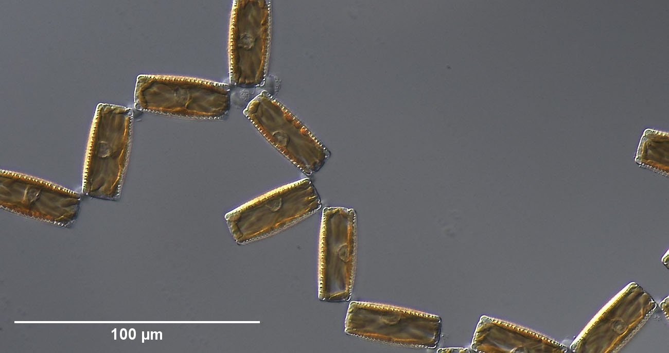
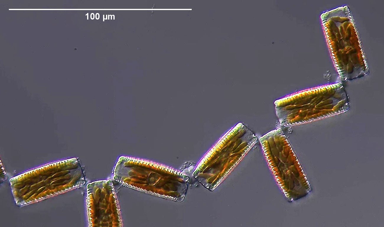
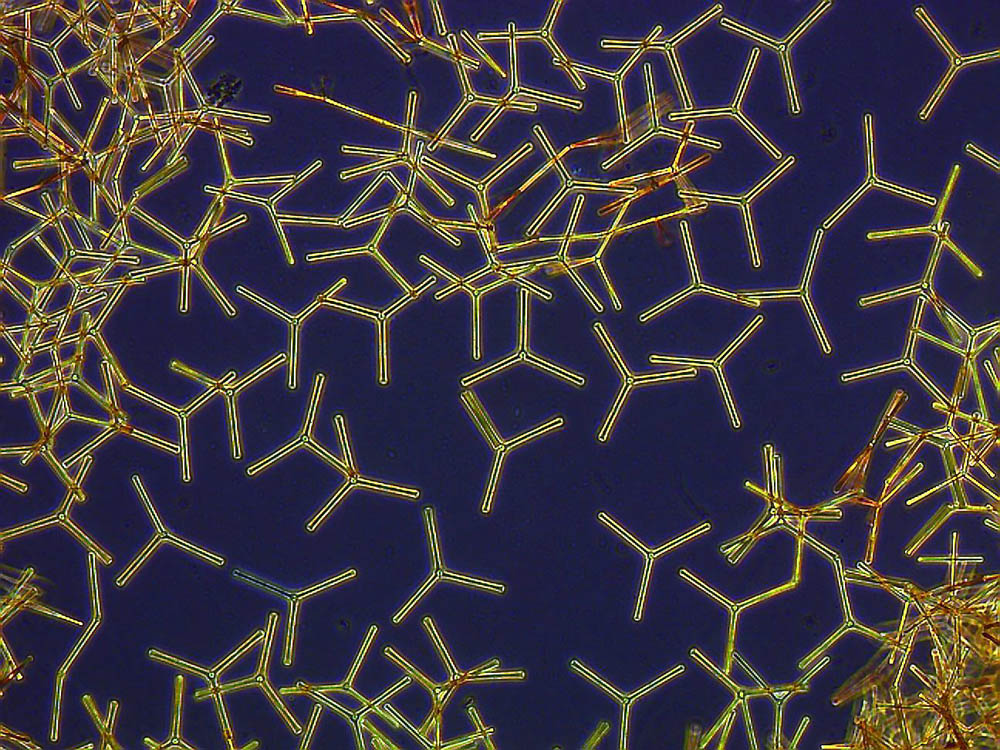
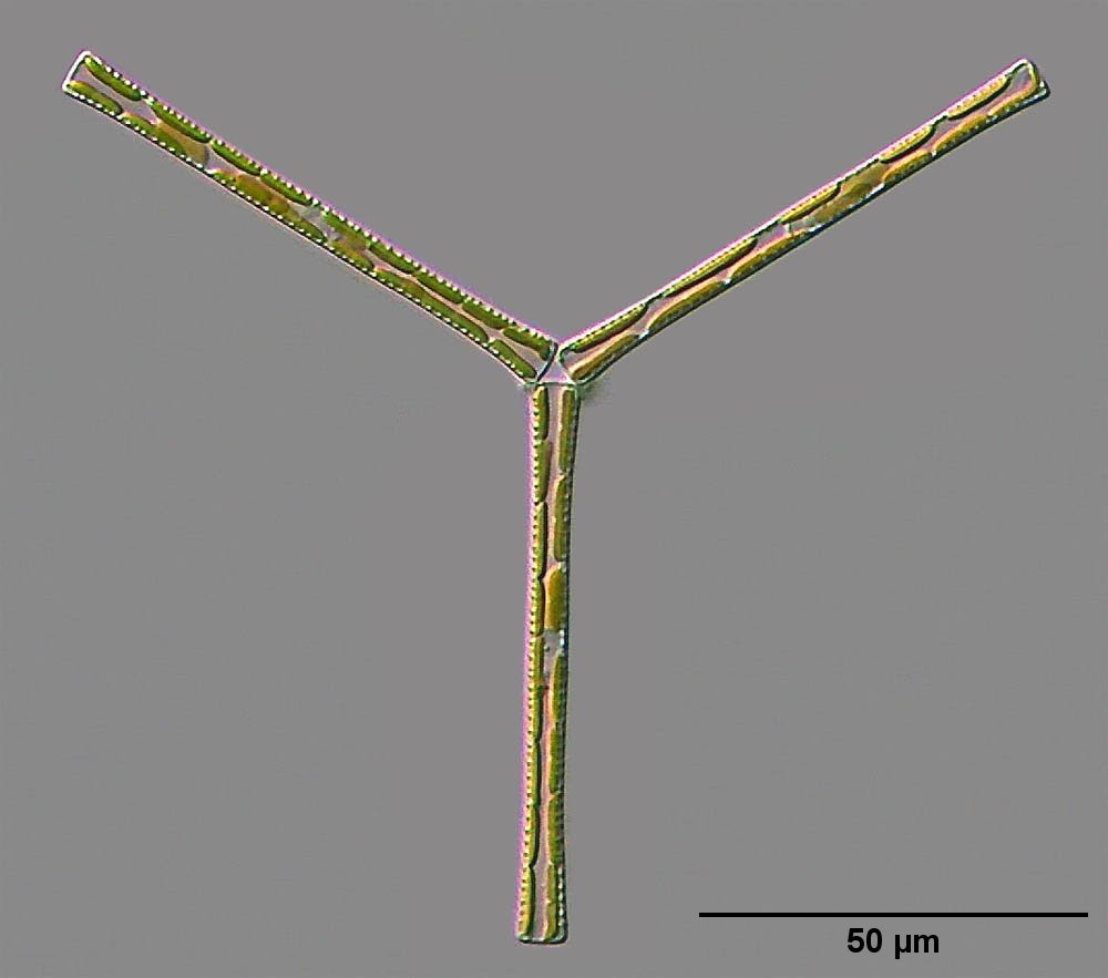
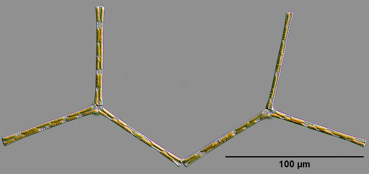
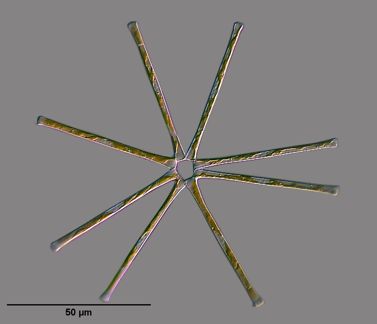
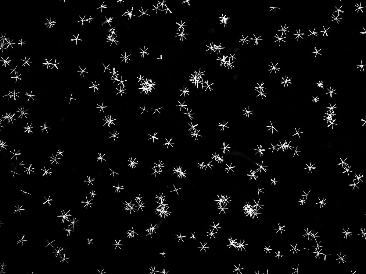
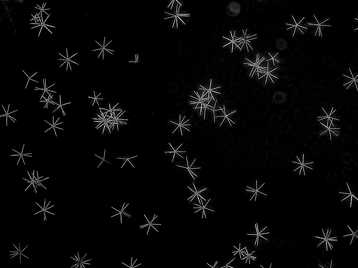
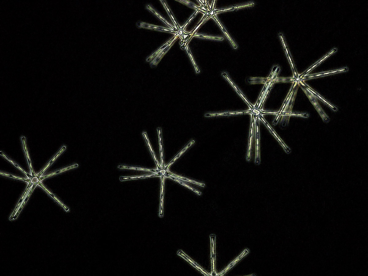
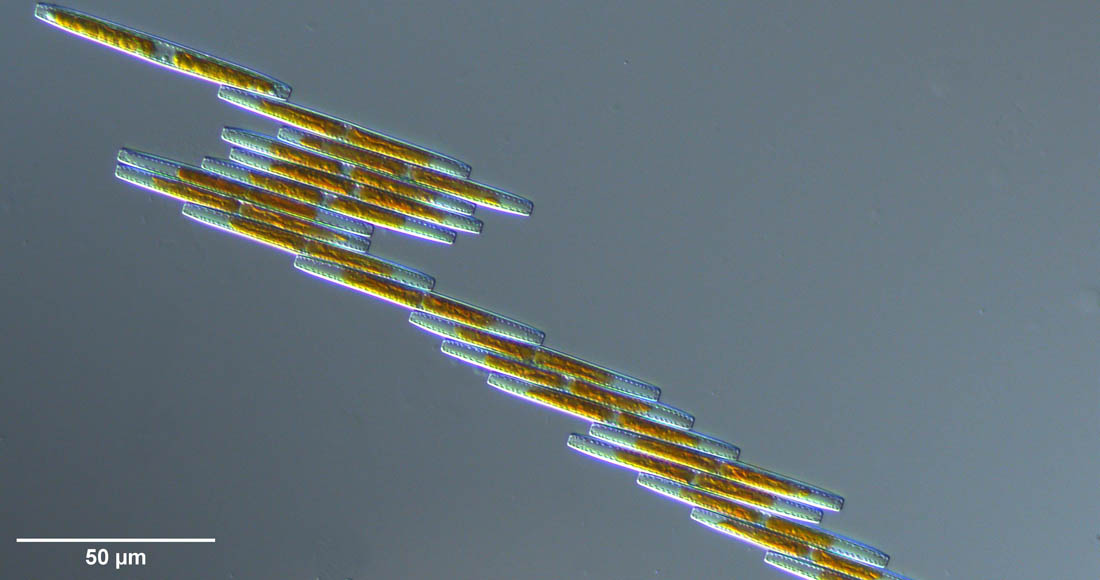
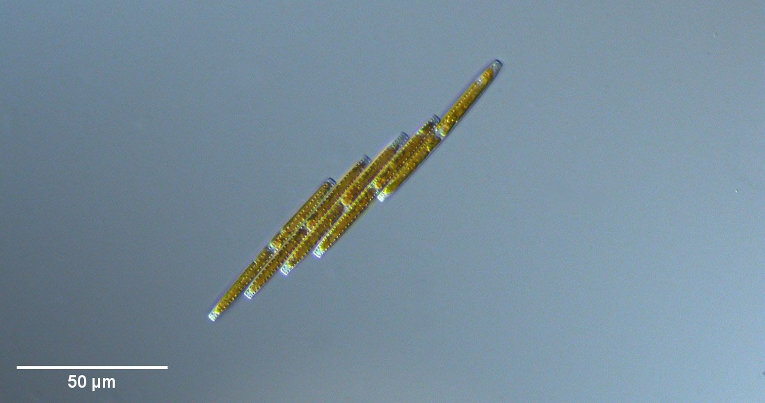
In the following image gallery some images of the cultures of Diatoma tenuis are shown. Longer chain-like structures, as well as short planktonic colony forms, are observed in this species. In culture, structures of one star formed by three diatoms and structures of up to four such connected stars occurred. In addition, there are transitional forms.
This video shows the sedimentation of Diatoma tenuis colonies in a Petri dish in 200-fold time lapse. The focus was on the bottom of the dish.
With Diatoma ehrenbergii another species of the genus Diatoma could be kept in culture. As shown in the images of the image gallery on the lower left, chain-like colonies, which adhere to the substrate, occur in coexistence with planktonic, mostly stellate colonies. Thereby, the short planktonic colonies are often formed by separation from long sessile colonies.
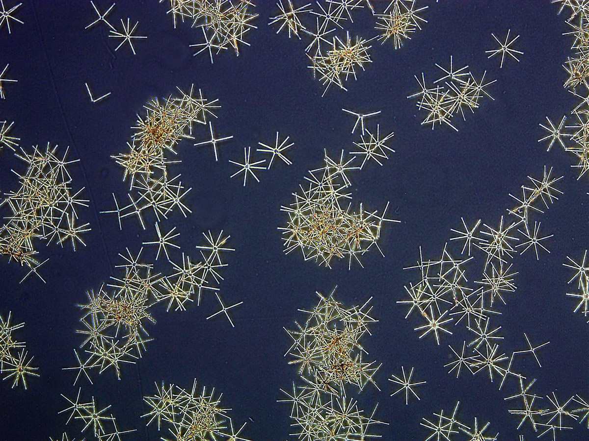
Below are images of cultures of Asterionella formosa. All cultivated planktonic species tended to multiply rapidly and reached high densities.
As a final example of chain-shaped colonies, a image gallery and a video of Bacillaria paxillifera (Bacillaria paradoxa) is shown below.
The diatoms have a raphe and exhibit a remarkable mobility, which is apparent in parallel displacement of the diatoms relative to neighbouring diatoms.
Dr. Nisch has also taken SEM images of Bacillaria paxillifera, which can be seen in an image gallery below. One can easily recognize the EPS, which serve the relative displacement. The preparation corresponds to that of the Diatoma species. Since thereby the samples were dried in the air after dehydration, artifacts are to be expected.
In nature, large colonies are easily broken into fragments by external influences such as a turbulent flow, so that only small colonies usually are found. Fragments can easily be drifted and may help spread.