In this video, you can see a region of a colony of Frustulia vulgaris living in tubes, in 50x time-lapse. This diatom builds and inhabits gelatinous tubes through which it moves. The video shows a region within a flat colony of a culture under differential interference contrast (DIC). In this region, the tubes lie one on top of the other without any apparent branches.
Frustulia vulgaris - Introduction and Structure of the Colonies
A number of pennate diatoms form tubes of extracellular polymeric substances (EPS) in which they live and move (for an introduction see [1]) and are therefore called tube-dwelling diatoms. This life form is mainly found in marine species, but also in some freshwater species. There are also diatoms that colonize tubes that have been made by other diatoms. In the sense of the term, these cohabitants are also tube-dwelling diatoms.
The subject of this work is the pennate diatom Frustulia vulgaris (Thwaites) De Toni 1891. It is a widespread tube-dwelling diatom found primarily in flowing waters. It thrives in freshwater and slightly brackish water. The studies presented here were based primarily on diatoms from the Neckar River (Germany 49°04'41.8 N 9°09'17.9 E) that were cultured.
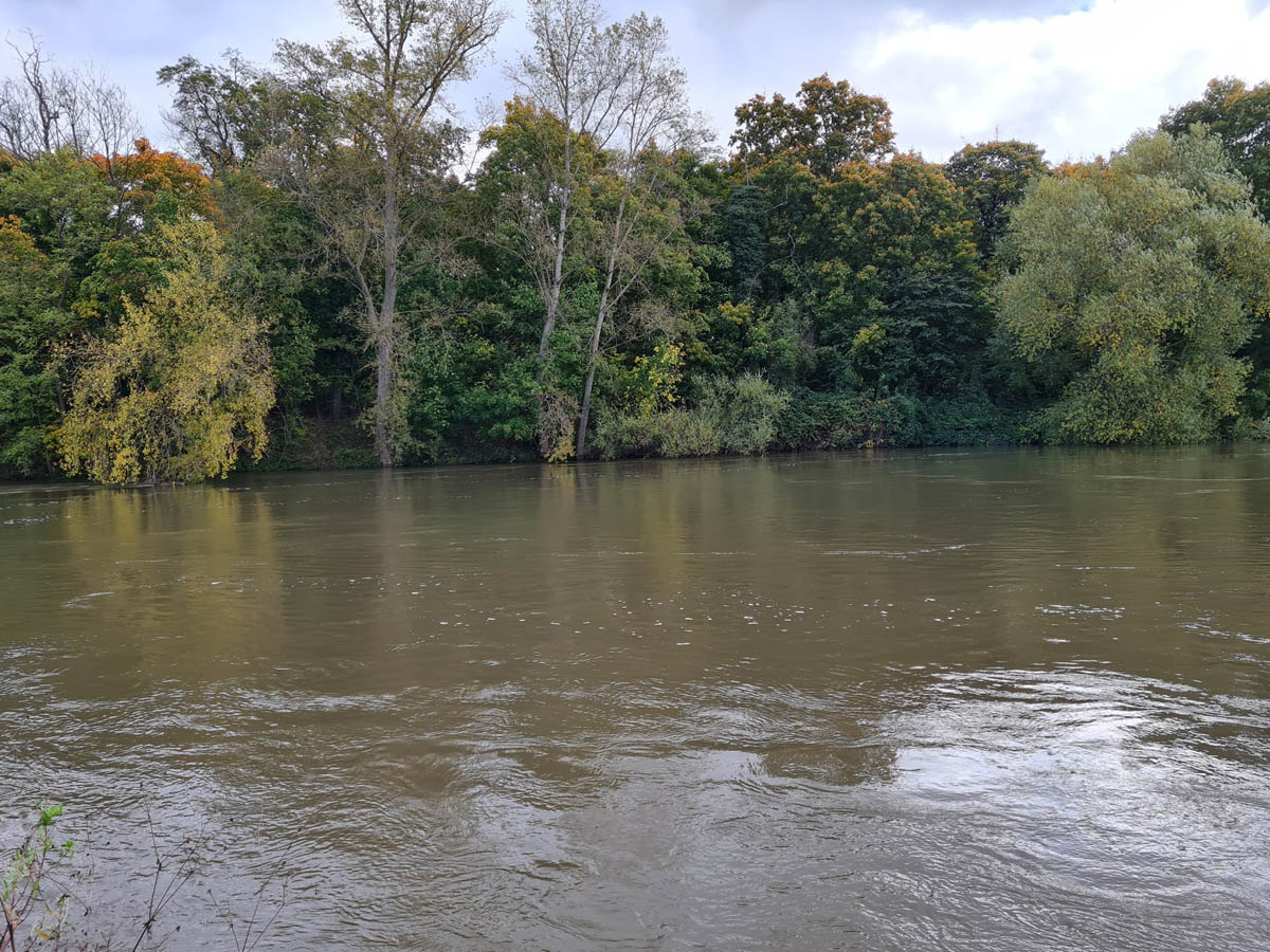 The Neckar River at the site of the finding with high water levels.
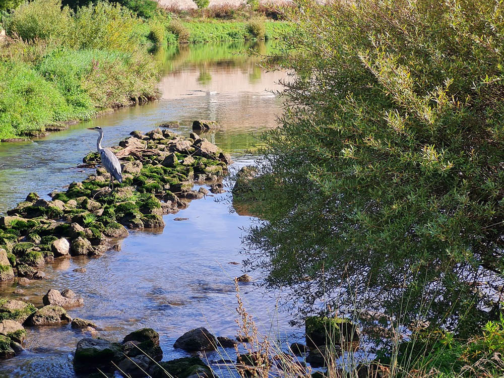 A heron is waiting for me at the confluence of the Zaber and Neckar rivers.
Sample of Frustulia vulgaris—colonies from a small stream in the Swabian-Franconian Forest in time-lapse (125x).
You can see two small F. vulgaris colonies attached to the substrate. Noteworthy are the amoebas, which appear to be hunting diatoms.
For unknown reasons, cultures of F. vulgaris isolated from this sample could only be maintained for a few generations.
Note on Cultivation
Cultivation using diatoms from the Neckar River was successful. Here are some key points on this topic (see the “Cultivation” section):
- Isolate a colony or parts of it using a fine pipette.
- As a rule, repeated purification by transferring the sample to clean water is necessary.
- Nutrient solution for phytoplankton (preferably f/2 according to Guillard)
- For batch cultures, regular “re-potting” approximately every two weeks
- Light exposure (fluorescent lamp) with day and night phases, each lasting about half a day
- Light phase with approx. 400 to 900 lx
- Cultivation in Petri dishes (polystyrene) allows for long-term observations.
Ecological Aspects of Tube-Dwelling Diatoms
The literature contains references to the ecological benefits of mucilage tubes in tube-dwelling diatoms. For these structures to be preserved, such benefits must outweigh the disadvantages.
Obvious disadvantages include:
- Energy expenditure for the production of building material and for activities related to the construction and maintenance of the tubes.
- Restriction to an effectively one-dimensional habitat. “Effectively” means that although the colonies have three-dimensional structures, the diatoms can only move in the direction of the tube. However, there are usually branches that represent choices at individual points. Depending on the colony’s shape, this restriction can make the search for light and nutrients more difficult.
- Restriction of migration to the colony’s location as long as the diatoms remain within the colony.
Advantages: Various advantages have been discussed. The literature specifically mentions (see, in particular, Drum [2] and Lobban [3] ):
- Protection from grazing, i.e., from predators. (see [4])
- Access to light and nutrients for colonial forms that rise above the substrate. Ability to rotate around the apical axis to optimize light uptake.
- Protection from disease (1)
- Protection from drastic changes in salinity in marine species (2)
- Protection against drying out (3)
(1) Young F. vulgaris—colonies are closed to the outside and may be axenic.
(2) However, the colony must be highly permeable to nutrients and metabolic products.
(3) A thick layer of colonies—for example, on a rock—dries slowly. A single thin colony does not provide effective protection in the case of F. vulgaris. As our observations show, the tubes collapse when they dry out, and the diatoms die quickly.
Access to light appears to be a disadvantage in connection with restricted movement, but also an advantage in the case of large colonies. Whether it is an advantage or a disadvantage may vary from case to case.
Protection against predators can be observed both in the wild and in laboratory samples. In the aforementioned stream sample, amoebas were found that could successfully engulf individual diatoms by phagocytosis, but not F. vulgaris colonies. The following two videos demonstrate this.
Time-lapse view (250x speed) of a sample from a stream in the Swabian-Franconian Forest. An amoeba can be seen engulfing diatoms.
An amoeba in the same sample attempting to engulf a F. vulgaris colony, shown in time-lapse (30x speed). The attempted phagocytosis is repeatedly abandoned without success.
Shape of the colonies
Colonies of the same species can vary greatly. G.H.K. Thwaites first described this diatom and its colonies. In his publication
Further observations on the Diatomaceae with descriptions of new genera and species. Annals and Magazine of Natural History, Series 2 1, 161-172
Thwaites notes:
“The species is most abundant in shallow streams, covering stones, &c. with a dark brown gelatinous coating, but in which a linear arrangement of the frustules may frequently be detected. When the plant occurs in deeper water, the ordinary Schizonema filaments make their appearance, which are much-branched when growing in rapid streams, but when occurring in still water, or where there is only a slight current, are simple or nearly so.”
Schizonema vulgare is the basionym of Frustulia vulgaris. Thwaites' drawing of a colony is shown below.
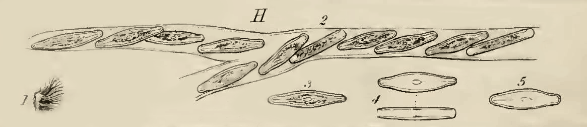 Illustration of a Frustulia vulgaris colony with branching, according to Thwaites, G.H.K. (1848)
As described by Krammer, K. and Lange-Bertalot, H. (1986), F. vulgaris colonies occur both aerially on wet rocks and planktonically as free-floating gelatinous tubes.
In Petri dishes, it colonizes the substrate with a network of tubes that exhibit branching. The following images were taken using a stereomicroscope and show a young colony. Only a few branches are visible.
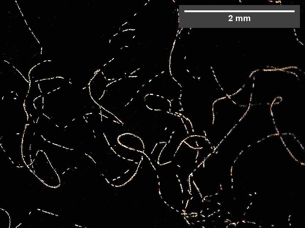 Overview image of a colony taken with a stereo microscope in dark-field imaging.
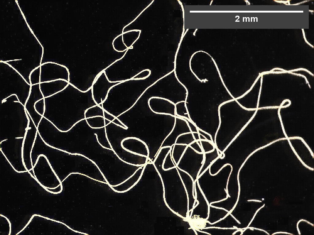 Superimposition of images over a one-hour period.
The situation is similar to growth on flat stones, where slimy colonies can be observed.
If a cuvette containing three-dimensional wire structures is inoculated, these structures become colonized. The following two images show such a cuvette.
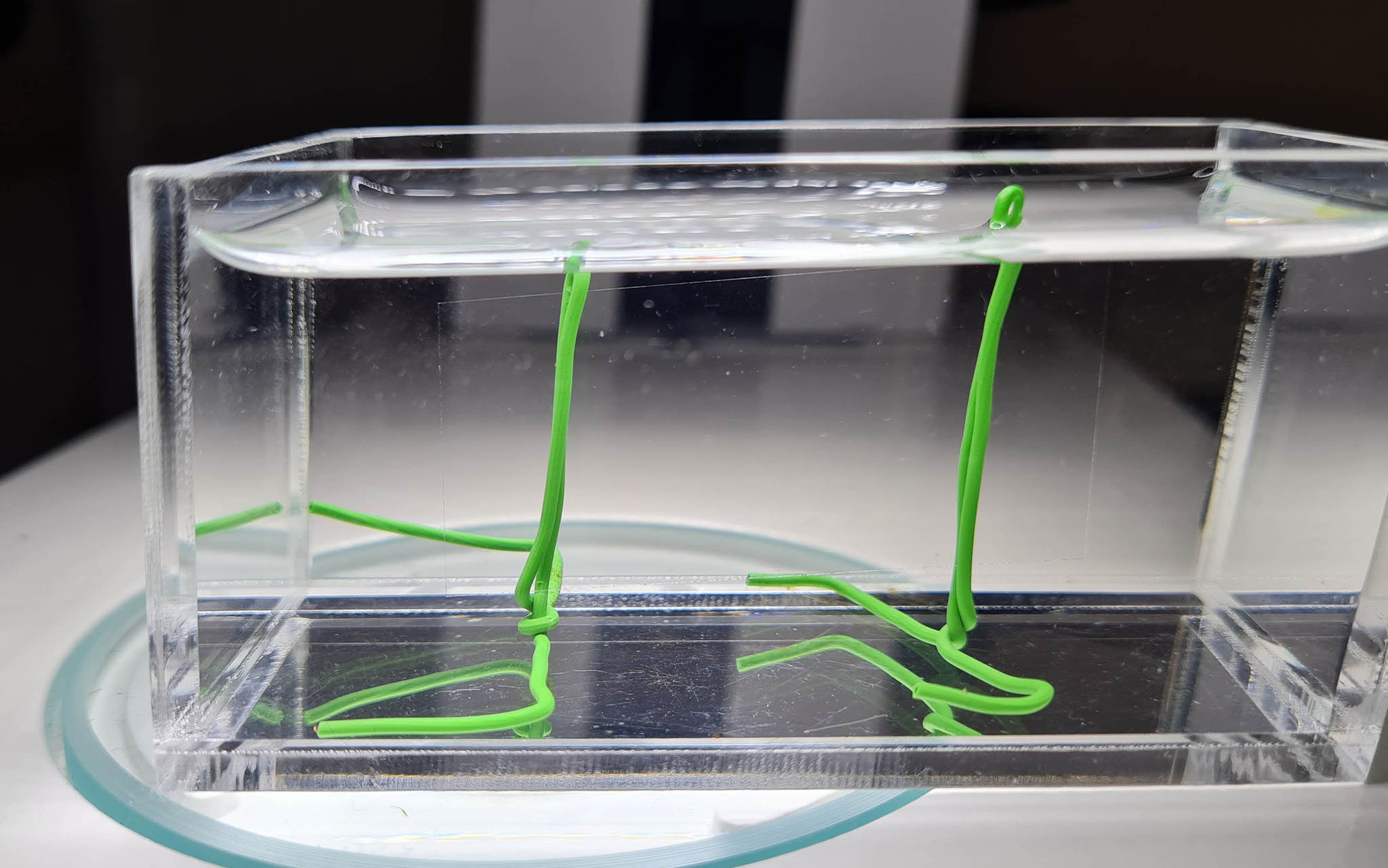 A simple structure consisting of coated wire and a large vertical cover glass, which was inserted into a cuvette.
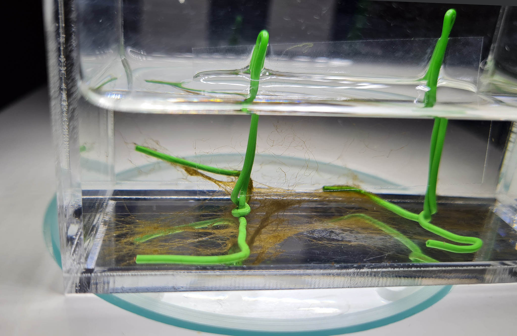 The cover slip is colonized from below via the stand. A uniform network forms there, similar to the colonization of a Petri dish.
After three to four weeks, filaments began forming and bridging the open spaces. These filaments then developed into net-like structures, with parts of the colonies that had detached from the substrate also contributing to this process. The following videos were recorded using a stereomicroscope with a horizontal optical axis. The green wire has a diameter of 0.8 mm.
Network-like spatial structures form. Recorded with a stereomicroscope using brightfield and darkfield modes with a horizontal viewing angle (500x time-lapse).
The colonized wire, viewed under a stereomicroscope (250x time-lapse).
Extensions of the colony protrude beyond the wire and move.
Although long filaments extending into space could not be observed, short filaments, as seen in videos of the wire being colonised, were visible. This is probably because there is no water current. Rather than developing into long filaments, they adhere to existing structures at their ends. The origin of these short filaments, which move actively, will be discussed in subsequent articles.
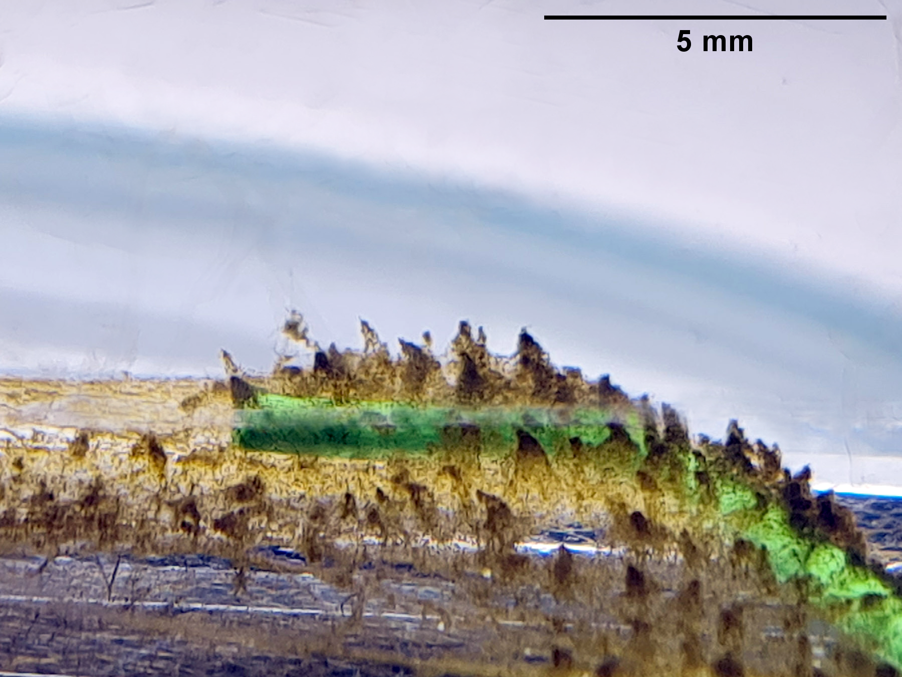
In the densest culture reached, pyramid-like structures can be seen along a coated wire and in the surrounding area of this older culture. The image was taken with a horizontal direction of view. The height of these structures is on the order of the wire's thickness, i.e., 0.8 mm.
Even on a flat surface such as a Petri dish, raised structures can be seen in older cultures. However, these are not arranged in a regular pattern. After they have developed for some time, one can find many individual, freely moving diatoms beneath them.
Selected publications:
[1] Round, F.E., Crawford, R.M. and Mann, D.G. (1990) The diatoms: biology and morphology of the genera. Cambridge University Press, Cambridge.
[2] Drum, R.W. (1969) Light and electron microscope observations on the tube‐dwelling diatom Amphipleura rutilans (Trentepohl) Cleve. Journal of Phycology 5(1), 21-26, doi: 10.1111/j.1529-8817.1969.tb02570.x.
[3] Lobban, C.S. (1989) Environmental factors, plant responses, and colony growth in relation to tube-dwelling diatom blooms in the Bay of Fundy, Canada, with a review of the biology of tube-dwelling diatoms. Diatom research 4(1), 89-109, doi: 10.1080/0269249X.1989.9705055.
[4] Schaeffer, A. A. (1916) On the feeding habits of ameba. Journal of Experimental Zoology 20(4), 529-584.
More articles on F. vulgaris are planned.